MRI Lumbar Spine
Last updated September 03, 2026
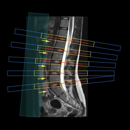
Protocol sequences
| Localizer_3_planes |
| I. Myelogram_Coronal_Spine |
| II. T2_Sagittal_Spine |
| III. T1_Sagittal_Spine |
| IV. STIR_Sagittal_Spine |
| V. T2_Axial_Disk |
| VI. T1_Axial_Disk |
Introduction
MRI Lumbar spine is one of the common scans in MRI. Scan is mainly requested for patients who suffer from backaches due to intervertebral disk bulging. Five to six sequences are performed routinely, but protocol may occasionally change due to pathologies including metastasis, fractures and scoliosis.
Patient preparation
- Ask the patient to remove metallic items such as jewellery and remove any clothing that contains ferromagnetic materials.
- Obtain MR safety consent after evaluating possible MR contraindications and take the necessary action if required.
- Explain that gradient noise and couch movements are normal.
- Demonstrate how to use the nurse call system and explain when to use it.
- Explain the importance of remaining still during the scan.
- Help claustrophobic patients overcome their fear through demonstrations, or allow a companion to stay with the patient during the scan.
- Inform the patient of the expected duration of the scan.
Patient positioning
- Posterior coil, which is in the scanner couch, is used for the scan.
- Position the patient head first and supine.
- Make sure that the patient lies centrally and symmetrically in the MR couch.
- Provide a cushion under the knees for extra comfort and reassure the patient.
- Position the patient’s limbs without forming body loops.
Explanation: Body loops may generate eddy currents, which can cause RF burns on the skin. Insulating soft pads may be used to break conductive loops that may form between the feet and thighs or between the trunk and the arm.
- Provide ear plugs to reduce scanner noise.
- Center the scan on the Iliac crest.
Sequences in detail
I. Myelogram_Coronal_Spine
Planning
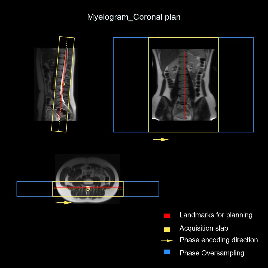
- Scan coverage: Include the entire spinal canal of the lumbar spine.
- Plan block angulation: Align to the spinal canal.
Parameters
FOV (cm)33x33
TR3200
Matrix352x352
TE500
No Wrap2x1
NAQ1
Slices1
Phase DirectionR to L
Slice thickness (mm)40
FLIP90
Gap (mm)0
Purpose
- To view the spinal canal for planning and anatomical evaluation.
II. T2_Sagittal_Spine
Planning
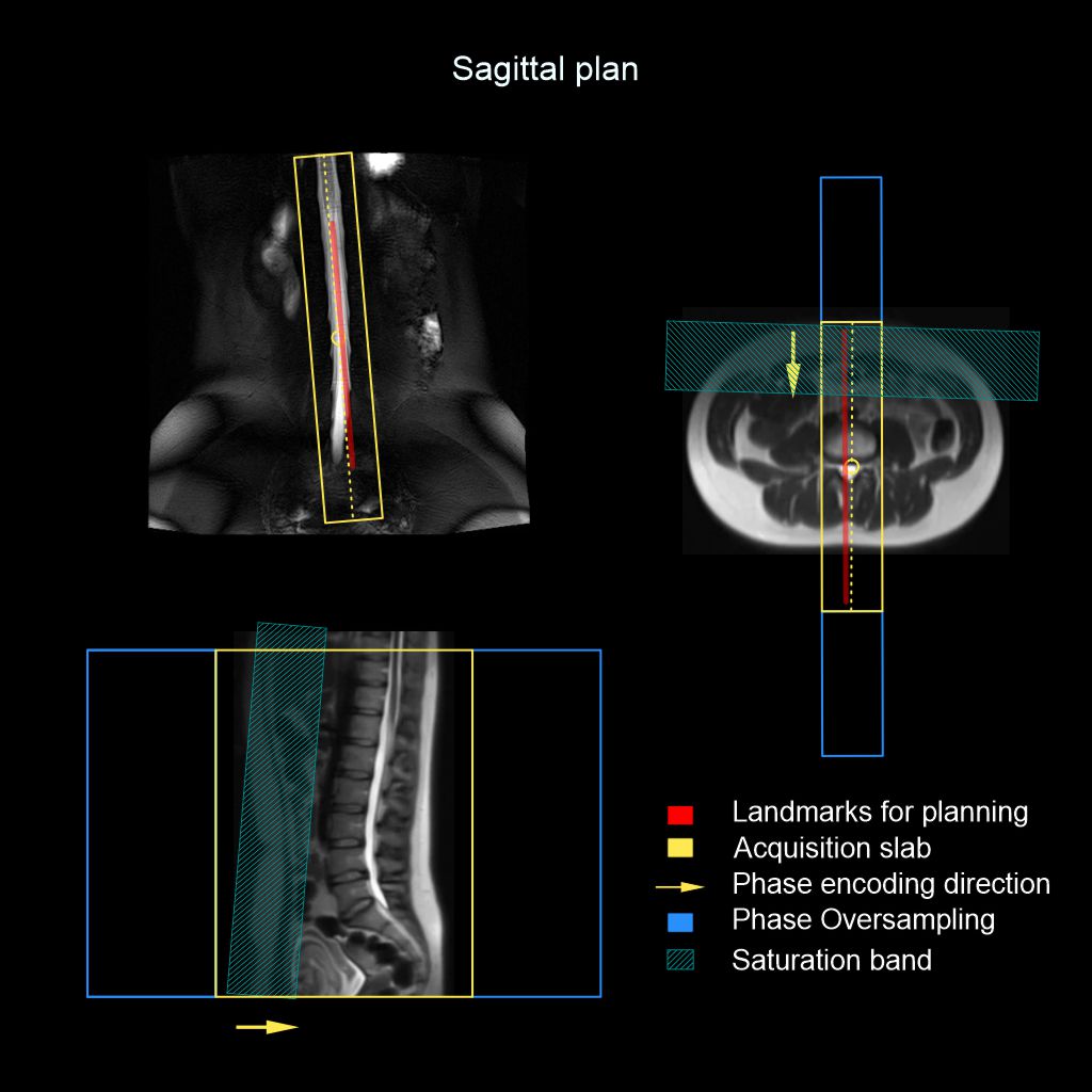
- Scan coverage: Include the entire lumbar spine, entire neural foramina and immediate paraspinal soft tissue. FOV should cover from T12 to S1.
- Plan block angulation: Align the sagittal slices parallel to the spinal canal in the coronal myelogram.
- Technical applications: Place a saturation band to cover the abdominal wall and bowel. This minimizes motion artifacts due to bowel and abdominal wall movements.
Parameters
FOV (cm)33x30
TR2919
Matrix352x352
TE121.5
No Wrap2x1
NAQ3
Slices11
Phase DirectionF to H
Slice thickness (mm)4
FLIP90
Gap (mm)0.9
Purpose
- Anatomical evaluation.
III. T1_Sagittal_Spine
Planning
- Scan coverage: Include the entire lumbar spine, entire neural foramina and immediate paraspinal soft tissue. FOV should cover from T12 to S1.
- Plan block angulation: Align the sagittal slices parallel to the spinal canal in the coronal myelogram.
- Technical applications: Place a saturation band to cover the abdominal wall and bowel. This minimizes motion artifacts due to bowel and abdominal wall movements.
Parameters
FOV (cm)33x30
TR570
Matrix320x320
TE13.5
No Wrap2x1
NAQ2
Slices11
Phase DirectionF to H
Slice thickness (mm)4
FLIP90
Gap (mm)0.9
Purpose
- Evaluate spinal bone marrow.
IV. STIR_Sagittal_Spine
Planning
- Scan coverage: Should include the entire lumbar spine, entire neural foramina and immediate paraspinal soft tissue. FOV should cover from T12 to S1.
- Plan block angulation: Align the sagittal slices parallel to the spinal canal in the coronal myelogram.
- Technical applications: Place a saturation band to cover the abdominal wall and bowel. This minimizes motion artifacts due to bowel and abdominal wall movements.
Parameters
FOV (cm)33x30
TR3360
Matrix256x256
TE60
No Wrap2x1
NAQ3
Slices11
Phase DirectionF to H
Slice thickness (mm)4
FLIP120
Gap (mm)0.9
TI140
Purpose
- Differentiate focal lesions.
V. T2_Axial_Disk
Planning
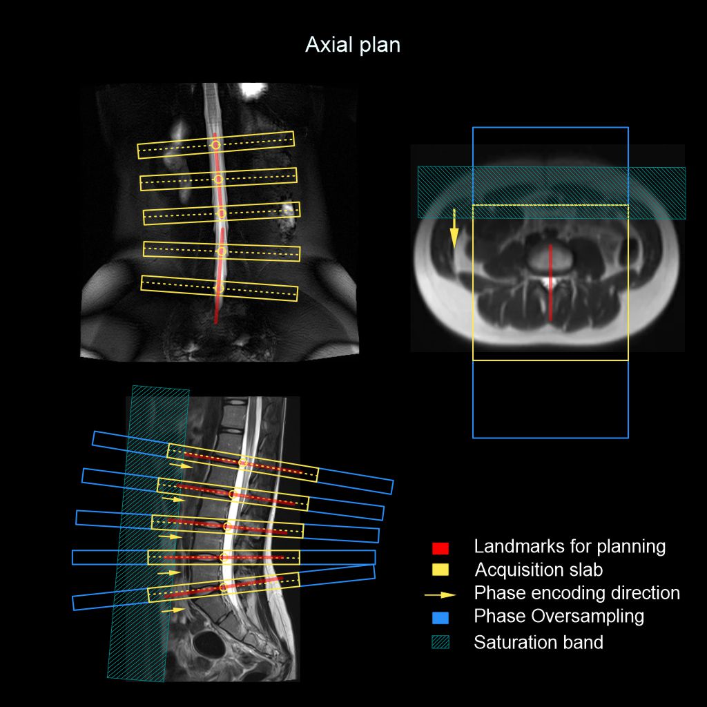
- Scan coverage: Include the spinal canal and the neural foramina.
- Plan block angulation: Align axial slices perpendicular to the spinal canal in the coronal myelogram, and parallel to the intervertebral disks in the sagittal T2 image.
- Technical applications: Place a saturation band to cover the abdominal wall and bowel. This minimizes motion artifacts due to bowel and abdominal wall movements.
Parameters
FOV (cm)20x20
TR3729
Matrix224x224
TE119
No Wrap2x1
NAQ4
Slices15
Phase DirectionA to P
Slice thickness (mm)4
FLIP90
Gap (mm)0.8
Purpose
- Anatomical evaluation and diagnosis of disk bulging.
VI. T1_Axial_Disk
Planning
- Scan coverage: Include the spinal canal and the neural foramina.
- Plan block angulation: Align axial slices perpendicular to the spinal canal in the coronal myelogram, and parallel to the intervertebral disks in the sagittal T2 image.
- Technical applications: Place a saturation band to cover the abdominal wall and bowel. This minimizes motion artifacts due to bowel and abdominal wall movements.
Parameters
FOV (cm)20x20
TR475
Matrix256x256
TE20
No Wrap2x1
NAQ2.5
Slices15
Phase DirectionA to P
Slice thickness (mm)4
FLIP90
Gap (mm)0.8
Purpose
- Anatomical evaluation.
Reference
- Douglas N. Mintz, MD, FACR, Chair, Saad Ali, MD, Avneesh Chhabra, MD, and Thomas Lee, MD. 2023. ACR–ASNR–SABI–SSR Practice parameter for the performance of magnetic resonance imaging (MRI) of the adult spine.
- Sollmann N, Fields AJ, O'Neill C, Nardo L, Majumdar S, Chin CT, Tosun D, Han M, Vu AT, Ozhinsky E, Shah LM, Harris RE, Lobo R, Anderst W, Herzog R, Psioda MA,Standaert CJ, Price RT, Lotz JC, Link TM, Krug R. Magnetic Resonance Imaging of the Lumbar Spine: Recommendations for Acquisition and Image Evaluation from the BACPAC Spine Imaging Working Group. Pain Med. 2023 Aug 4;24(Suppl 1):S81-S94. doi: 10.1093/pm/pnac130. PMID: 36069660; PMCID: PMC10403314.
- Zerunian M, Pucciarelli F, Caruso D, De Santis D, Polici M, Masci B, Nacci I, Del Gaudio A, Argento G, Redler A, Laghi A.Fast high-quality MRI protocol of the lumbar spine with deep learning-based algorithm: an image quality and scanning time comparison with standard protocol. Skeletal Radiol. 2024 Jan;53(1):151-159. doi: 10.1007/s00256-023-04390-9. Epub 2023 Jun 28. PMID: 37369725; PMCID: PMC10661795.
- Sayah A, Jay AK, Toaff JS, Makariou EV, Berkowitz F. Effectiveness of a Rapid Lumbar Spine MRI Protocol Using 3D T2-Weighted SPACE Imaging Versus a Standard Protocol for Evaluation of Degenerative Changes of the Lumbar Spine. AJR Am J Roentgenol. 2016 Sep;207(3):614-20. doi: 10.2214/AJR.15.15764. Epub 2016 Jun 8. PMID: 27275868.