CT Myelography (CTM)
Last updated June 27, 2026
By Radiohelp Staff
Similar expressions
CT myelography/ CT myelogram/ CECT myelogram/ CTM
Introduction
Myelo-CT is used to assess pathologies related to the spinal cannel, and commonly conducted for patients who are contraindicated for MRI scans. CTM can be used to diagnose pathologies such as dorsal thoracic web, spinal arachnoid cyst and CSF leaks. Reach CT Cisternography section for CSF leak assessment.
Patient preparation
Lumbar puncture is performed by an experienced physician, and iodinated contrast media is injected into the spinal sub-arachnoid space.
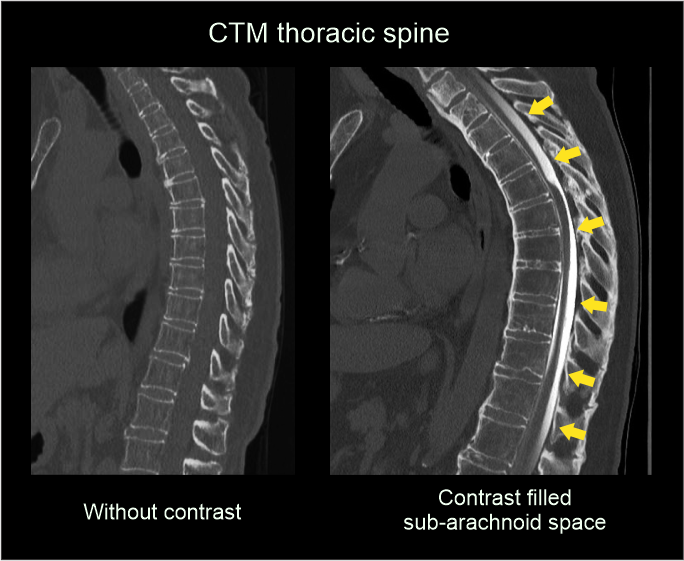
- Opacified contrast, with aid of a tilting table, is moved to the spinal sub-arachnoid space of the desired region (lumber, thoracic or cervical).
Explanation: opacified CSF can be moved from a higher point to a lower point by creating a slope.
- Patient is rolled from side to side.
Explanation: this is to spread contrast media throughout the sub-arachnoid space.
To get the best opacified region, imaging area needs to be at a lower point while the rest is raised. Following positions can be used to direct contrast into the needed region.
Cervical spine myelogram.
- Position in prone.
- Place hands either side of the body.
- Hyper extend the neck, and slightly raise pelvic and chest areas.
Thoracic spine myelogram.
- Position in supine.
- Slightly raise head and pelvic region by placing pads under them.
Lumbar sacral myelogram.
- Position in supine.
- Slightly raise head and chest areas.
Patient positioning
Patient can be positioned for imaging, ones the needed region is opacified with contrast media. Use lateral scout image to pre assess contrast position.
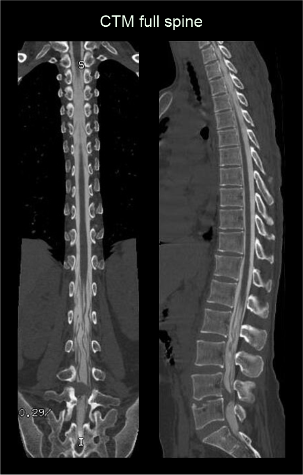
- Patient positioning for imaging is same as in CT spine examinations. Please refer: cervical spine, thoracic spine, lumbar sacral spine and full spine.
Scan planning
- Scan planning is same as in CT spine examinations. Please refer: cervical spine, thoracic spine, lumbar sacral spine and full spine.
Post-processing
- Sagittal and coronal images with ≤ 2 mm slice thickness in bone window (WW: 3500, WL: 350).
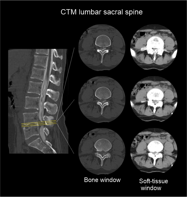
- Contagious axial slices in bone window and soft-tissue window (WW:500, WL:50) with ≤ 2mm and ≤ 3mm slice thicknesses respectively.
- Axial images through inter-vertebral disk for the assessment of spinal cord compressions.
Reference
- Amrhein, Timothy J MD, Chair, Goldman-Yassen, Adam MD, & Ali, Saad MD. (2024). ACR–ASNR–SPR practice parameter for the performance of myelography and cisternography. Retrieved from www.gravitas.acr.org.
- Wendl, C. M., Schambach, F., Zimmer, C., & Förschler, A. (2012). CT myelography for the planning and guidance of targeted epidural blood patches in patients with persistent spinal CSF leakage. AJNR. American journal of neuroradiology, 33(3), 541–544. https://doi.org/10.3174/ajnr.A2808
- Kranz PG, Luetmer PH, Diehn FE, Amrhein TJ, Tanpitukpongse TP, Gray L. Myelographic Techniques for the Detection of Spinal CSF Leaks in Spontaneous Intracranial Hypotension.AJR Am J Roentgenol. 2016 Jan;206(1):8-19. doi: 10.2214/AJR.15.14884. PMID: 26700332.
- Mamlouk MD, Shen PY, Dahlin BC. Modified Dynamic CT Myelography for Type 1 and 2 CSF Leaks: A Procedural Approach.AJNR Am J Neuroradiol. 2023 Mar;44(3):341-346. doi: 10.3174/ajnr.A7784. Epub 2023 Feb 2. PMID: 36732032; PMCID: PMC10187812.
- Boto J, Vargas MI. A new modified technique of dynamic CT myelography to detect dural tears in spontaneous intracranial hypotension.J Neuroradiol. 2024 Mar;51(2):210-213. doi: 10.1016/j.neurad.2023.07.004. Epub 2023 Jul 26. PMID: 37499791.
- Kranz PG, Amrhein TJ, Gray L. CSF Venous Fistulas in Spontaneous Intracranial Hypotension: Imaging Characteristics on Dynamic and CT Myelography.AJR Am J Roentgenol. 2017 Dec;209(6):1360-1366. doi: 10.2214/AJR.17.18351. Epub 2017 Oct 12. PMID: 29023155.