CT Renal Angiogram
Last updated July 01, 2026
By Radiohelp Staff
Similar expressions
CECT renal angio/ CT renal angiogram/ CECT renal arteries
Introduction
CT Renal Angiogram is used to diagnose conditions related to the renal arteries including aneurysms, arterial stenosis, congenital abnormalities, neoplasms and kidney transplant evaluation.
Patient preparation
- Explain the procedure clearly and kindly.
- Check contraindications for contrast media administration and radiation exposure.
- Remove metals related to the interested region.
- Place an Intra venous (IV) cannula in a stable vein of an arm – green-18G cannula.
Explanation: green cannula has a higher lumen diameter, which can withstand higher flow rate.
- Provide 1000ml of water to drink within 20-30min without emptying the urinary bladder.
Explanation: this is to adequately fill the bladder with urine, which promotes visualization of ureters.
- Start the scan when the patient is having a fully filled bladder.
- Explain and practice breath in and hold technique.
Explanation: avoids motion un-sharpness of organs and blood vessels.
Patient positioning
- Position the patient in supine and feet first on the imaging couch.
- Center the scanning area in the scanner iso-center [6].
Explanation: this reduces overall radiation exposure and increases image quality.
- Raise both hands above the head and place a positioning aide under hands.
Explanation: hands beside the trunk give streak artifacts and increase radiation exposure.
- Keep the arm with the IV cannula strait.
Explanation: to facilitate contrast flow.
- Plan the scan starting point at the nipple level.
Scan planning
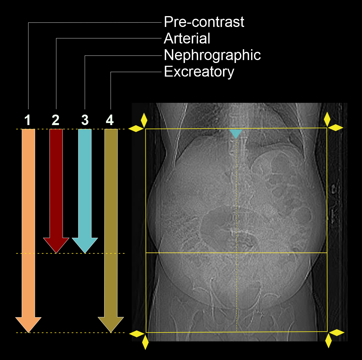
- Scan is performed under multiple phases. Usually, it has 4 phases.
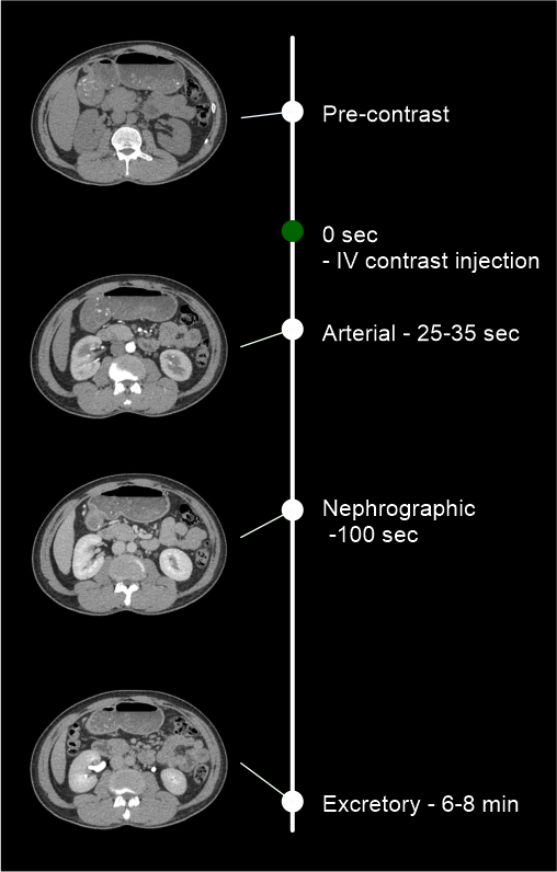
- Plan both pre-contrast and excretory phases to cover from dome of the diaphragm to a level just below the ischial tuberosities.
- Plan both arterial and nephrographic phases to cover form dome of the diaphragm to the upper margin of the sacroiliac joints.
Important: covering areas may be changed according to the patient condition and the radiologist’s preference.
- Set the phase initiation.
Explanation: Use bolus tracking method to start the angiographic phase on time. Keep a ten seconds gap between contrast initiation point and triggering point, and should not place a delay time to start the angiographic phase once the threshold value (180HU) is reached. Place scan-and-view slice (S and V) just above the diaphragm. Please refer abdominal aortogram scan for images.
IV contrast injection.
- Inject 80 ml (for a patient with 70-80kg body weight) of iodinated contrast media at a rate of 4.5-5ml/s.
- Inject 50-60ml of saline flush following the contrast injection.
Explanation: flushes remaining contrast in the veins of the injected hand, and maintains contrast flow for a longer time.
Important: experienced radiographer must be involved when administering IV contrast because injection rate, contrast volume, and technique might be changed according to the patient body weight, patient condition, scan capturing range, and CT scanner model.
Post-processing
- Multiplanar reconstruction (MPR) images for all phases in soft-tissue window (WW:500, WL:50) with slice thickness ≤ 5mm – refer to non-contrast abdomen scan for images.
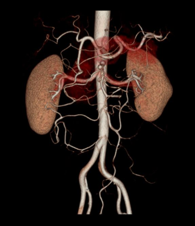
- 3d reconstruction of the renal arteries, abdominal aorta and common iliac arteries.
For kidney donors:
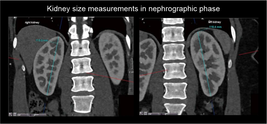
- Kidney size measurement in MPR images (Nephrographic phase).
- Renal artery pre-hilar arterial branching distance measurements on curved MPR or on 3D images, separately for both renal arteries.
- Renal artery diameter measurements from abdominal aorta to first branching on proximal, mid and distal points.
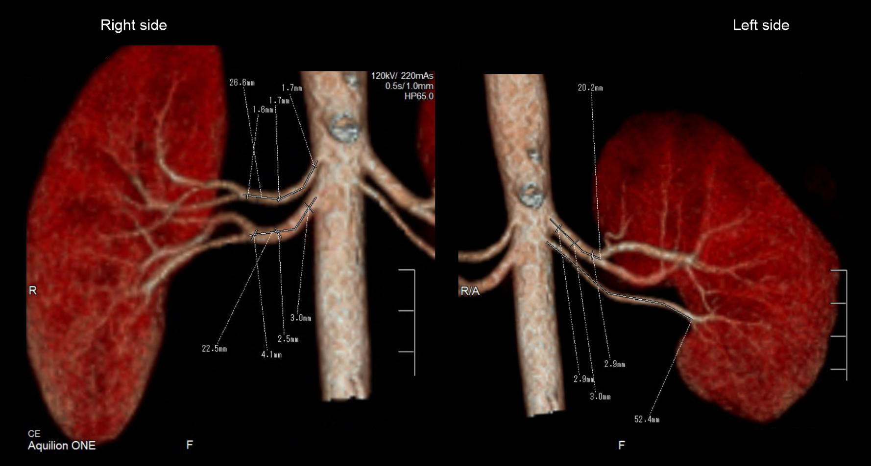
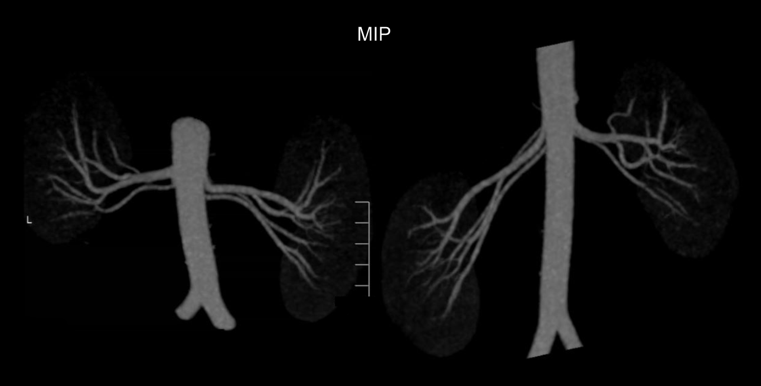
- MIP images through the kidneys to show small accessory renal arteries.
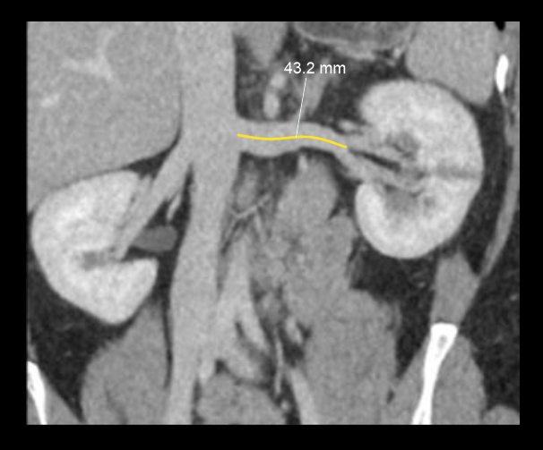
- Curved MPR images of nephrographic phase to show renal venous system and distance from inferior vena cava (IVC) margin to venous bifurcation.
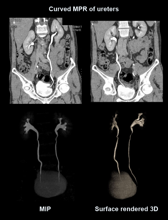
- 3D, MIP, curved MPR images from excretory phase to show ureters.
Reference
- Steven S. Raman, MD, Chair, Dorothy Gilbertson, MD, & Charles White, MD. (2021). ACR–NASCI–SIR–SPR Practice parameter for the performance and interpretation of body computed tomography angiography (CTA). Retrieved from www.gravitas.acr.org.
- Olga R. Brook, MD, Chair, Jessica Kurian MD, Alec Megibow, MD, MPH, FACR, & Michael Furman, MD. (2021). ACR–SABI–SAR–SPR practice parameter for the performance of computed tomography (CT) of the abdomen and computed tomography (CT) of the pelvis.Retrieved from www.gravitas.acr.org.
- Cho ES, Yu JS, Ahn JH, Kim JH, Chung JJ, Lee HK, Lee KH. CT angiography of the renal arteries: comparison of lower-tube-voltage CTA with moderate-concentration iodinated contrast material and conventional CTA.AJR Am J Roentgenol. 2012 Jul;199(1):96-102. doi: 10.2214/AJR.11.7450. PMID: 22733899.
- Hazırolan T, Öz M, Türkbey B, Karaosmanoğlu AD, Oğuz BS, Canyiğit M. CT angiography of the renal arteries and veins: normal anatomy and variants.Diagn Interv Radiol. 2011 Mar;17(1):67-73. doi: 10.4261/1305-3825.DIR.2902-09.1. Epub 2010 Feb 9. PMID: 20151356.
- Aghayev, A., Gupta, S., Dabiri, B. E., & Steigner, M. L. (2019). Vascular imaging in renal donors. Cardiovascular diagnosis and therapy, 9(Suppl 1), S116–S130. https://doi.org/10.21037/cdt.2018.11.02
- Romanyukha, A., Nzitunga, P. S., & Dolcet, A. (2022, April 28). CT patient positioning plays key role in radiation dose reduction.www.auntminnie.com.