CT Thoracic Spine
Last updated June 27, 2026
By Radiohelp Staff
Similar expressions
CT T spine
Introduction
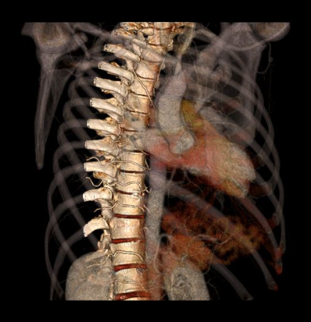
CT Thoracic spine protocol is mainly used to assess thoracic spine injures such as fractures. It also helps to diagnose spine neoplasms, congenital abnormalities and implant checks.
Patient preparation
- Explain the examination clearly and kindly.
- Ask to remove radio opaque items such as necklaces, underclothing and piercing related to the thoracic region.
Patient positioning
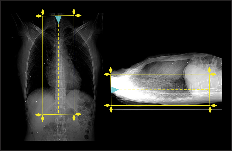
- Position in feet-first, supine and at the iso-center.
Explanation: patient in the iso-center reduces radiation exposure and improves image quality.
- Both hands are raised above the head.
Explanation: this avoids streak artifacts due to hands.
- Plan the scout start point at lower neck region and set scan direction outward the gantry.
Scan planning
- Plan the scan slab to cover from C7 to the first lumbar vertebra (L1).
- Reduce the field of view (FOV) as small as appropriate.
Explanation: smaller FOV increases the geometric resolution of the image.
Post-processing
- Sagittal and coronal images with ≤ 2 mm slice thickness in bone window (WW: 3500, WL: 350).
- Contagious axial slices in bone window and soft-tissue window (WW:500, WL:50) with ≤ 2mm and ≤ 3mm slice thicknesses respectively.
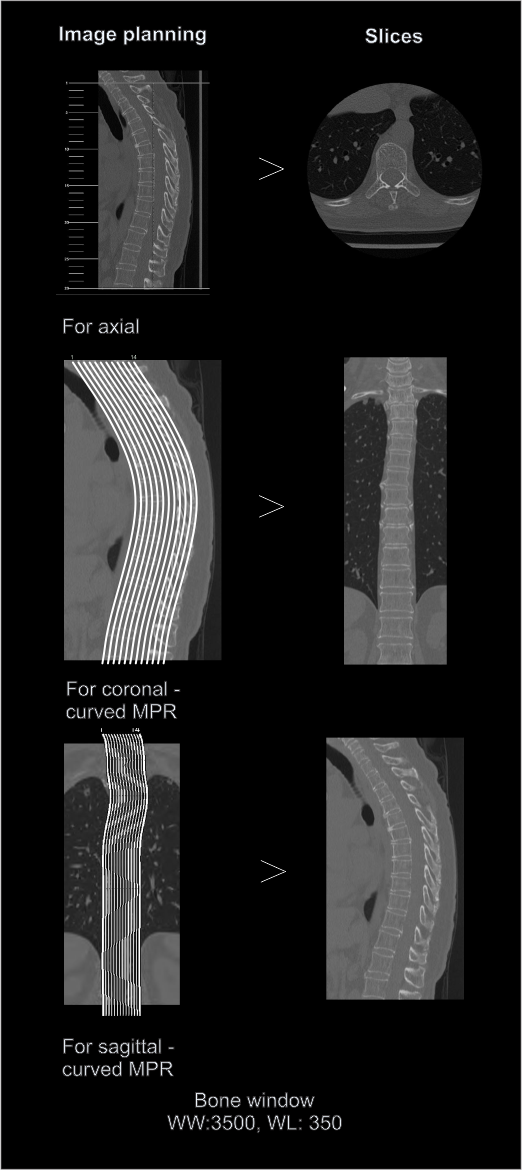
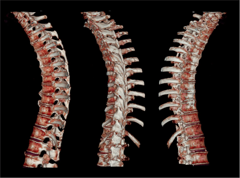
- Additionally, 3d images to show the thoracic-spine.
Reference
- Lubdha M. Shah, MD, Chair, Kristine A. Blackham, MD, & Kavita K. Erickson, MD. (2022). ACR–ASNR–ASSR–SPR practice parameter for the performance of computed tomography (CT) of the spine. Retrieved from www.gravitas.acr.org.
- Mannudeep K. S. Kalra, MD, Chair, Jessica Kurian MD, & Satinder Singh, MD, FSABI. (2023). ACR–SABI–SPR–STR Practice parameter for the performance of thoracic computed tomography (CT). Retrieved from www.gravitas.acr.org.
- Manca A, Chiara G, Bellizzi S, Valle P, Nicoli S, Campanella D, Regge D. Thoracic Spine CT Hidden Treasures: Lung Assessment and Extraspinal Findings in Patients with Vertebral Fractures Studied with Full FOV during Breath Hold: Technical Note. Tomography. 2022 Apr 2;8(2):999-1004. doi: 10.3390/tomography8020080. PMID: 35448714; PMCID: PMC9030083.
- Joaquim, A. F., & Patel, A. A. (2013). Thoracolumbar spine trauma: Evaluation and surgical decision-making. Journal of craniovertebral junction & spine, 4(1), 3–9. https://doi.org/10.4103/0974-8237.121616
- Gamanagatti, S., Rathinam, D., Rangarajan, K., Kumar, A., Farooque, K., & Sharma, V. (2015). Imaging evaluation of traumatic thoracolumbar spine injuries: Radiological review. World journal of radiology, 7(9), 253–265. https://doi.org/10.4329/wjr.v7.i9.253
- Romanyukha, A., Nzitunga, P. S., & Dolcet, A. (2022, April 28). CT patient positioning plays key role in radiation dose reduction.www.auntminnie.com.