CT Sinus
Last updated June 27, 2026
By Radiohelp Staff
Similar expressions
NCCT sinus/ CT sinus/ CT paranasal sinus
Introduction
CT Sinus is one of the routine scans and it helps to diagnose conditions such as sinusitis, fluid collections, nasal septum deviation, congenital abnormalities and tumors.
Patient preparation
- Remove hair clips, ear rings or any removable metal in the exposing area.
- Explain the procedure clearly.
- Ask the patient to be steady during the acquisition.
Patient positioning
There are two patient positioning methods as supine and prone.
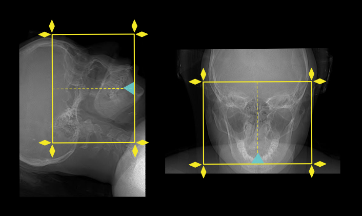
Method 1 - supine
- Position the patient in head first and supine on the scanner couch.
- Position the head in the head rest and use immobilizing bands.
- Tilt the head such that the line joining supra-orbital point and external auditory meatus (EAM) perpendicular to the floor, and ask to close the eyes (please find more information from our Brain non-contrast article).
Explanation: this method reduces the eye lens dose.
- Center the scanning area in the scanner iso-center [5].
Explanation: this reduces overall radiation exposure and increases image quality.
Method 2 - prone
- Position the patient in prone and head first, resting on the chin with hyperextended head.
Explanation: appearance of air-fluid level in supine and prone methods are different.
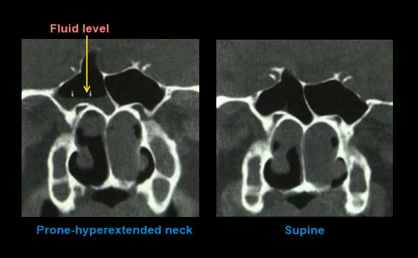
Doing the scan in both positions may necessary to differentiate lesions attached to the inner wall of the sinus because fluid levels change with the patient position, but not the attached neoplasms.
Scan planning
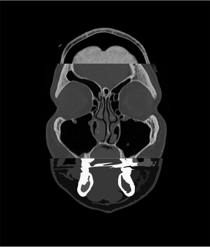
- In supine method, plan the scan slab to cover from the hard palate up to the superior border offrontal sinus.
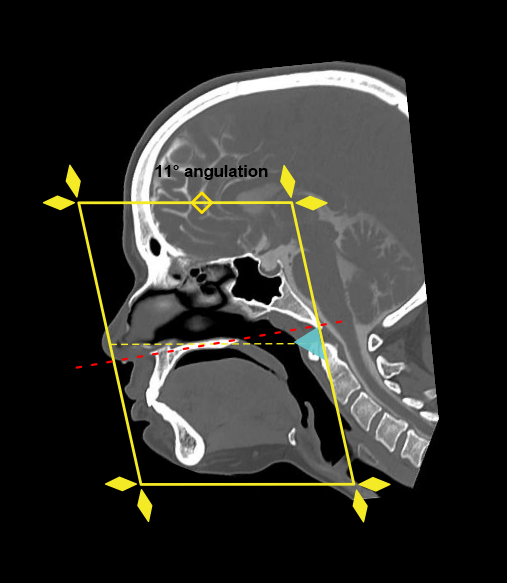
- In prone method (neck hyperextended), plan the scan slab to cover from nose to posterior margin of sphenoid sinus. Scan slab needs to be perpendicular to hard palate, which is achieved by tilting the gantry.
- Reducing the FOV to sinus area increases image quality [7].
Explanation: this increases geometric resolution.
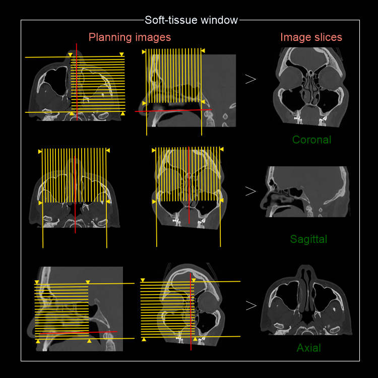
Post-processing
Axial, sagittal and coronal reformations with 2 mm thickness in bone (WW:4500, WL:450) and soft-tissue (WW:400, WL:40) window.
Reference
- Ashley H. Aiken, MD, Chair, Paul M. Bunch, MD, & Kavita K. Erickson, MD. (2021). ACR–ASNR–SPR Practice parameter for the performance of computed tomography (CT) of the extracranial head and neck.Retrieved from www.gravitas.acr.org.
- Babbel, R., Harnsberger, H. R., Nelson, B., Sonkens, J., & Hunt, S. (1991). Optimization of techniques in screening CT of the sinuses.AJNR. American journal of neuroradiology, 12(5), 849–854.
- Al Abduwani J, ZilinSkiene L, Colley S, Ahmed S. Cone beam CT paranasal sinuses versus standard multidetector and low dose multidetector CT studies. Am J Otolaryngol. 2016 Jan- eb;37(1):59-64. doi: 10.1016/j.amjoto.2015.08.002. Epub 2015 Aug 24. PMID: 26700263.
- Shpilberg KA, Daniel SC, Doshi AH, Lawson W, Som PM. CT of Anatomic Variants of the Paranasal Sinuses and Nasal Cavity: Poor Correlation With Radiologically Significant Rhinosinusitis but Importance in Surgical Planning. AJR Am J Roentgenol. 2015 Jun;204(6):1255-60. doi: 10.2214/AJR.14.13762. PMID: 26001236.
- Romanyukha, A., Nzitunga, P. S., & Dolcet, A. (2022, April 28). CT patient positioning plays key role in radiation dose reduction.www.auntminnie.com.
- Carberry, George & Lubner, Meghan & Wells, Shane & Hinshaw, J. (2016). Percutaneous biopsy in the abdomen and pelvis: a step-by-step approach.Abdominal radiology (New York). 41. 10.1007/s00261-016-0667-1.
- Salimova, N., Hinrichs, J. B., Gutberlet, M., Meyer, B. C., Wacker, F. K., & von Falck, C. (2022).The impact of the field of view (FOV) on image quality in MDCT angiography of the lower extremities. European radiology, 32(5), 2875–2882.https://doi.org/10.1007/s00330-021-08391-x