CT non-contrast Chest
Last updated June 03, 2026
By Radiohelp Staff
Similar expressions
CT chest/ CT non-contrast chest/ NCCT chest
Introduction
NCCT chest scan is routinely used to evaluate abnormalities discovered from other imaging modalities, including chest radiography (lung consolidations, nodules, trauma). This scan is combined with post contrast steps for the diagnosis of complex pathologies. Please reach our chest contrast, coronary angiography, pulmonary angiography and thoracic aortogram articles for more information.
Patient preparation
- Explain the procedure clearly and kindly.
- Remove metals related to the scanning area (underwear and necklaces).
- Check the suitability to expose radiation.
- Practice breathing technique – breath in and hold during the scan.
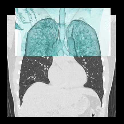
Patient positioning
- Position the patient in supine and feet first on the imaging couch.
- Place both hands above the head and place a positioning aide or a pillow under the hands for comfort.
Explanation: this reduces streak artifacts from the hands, and also avoids unnecessary radiation exposure to the hands.
- Center the scanning area in the scanner iso-center [6].
Explanation: this reduces overall radiation exposure and increases image quality.
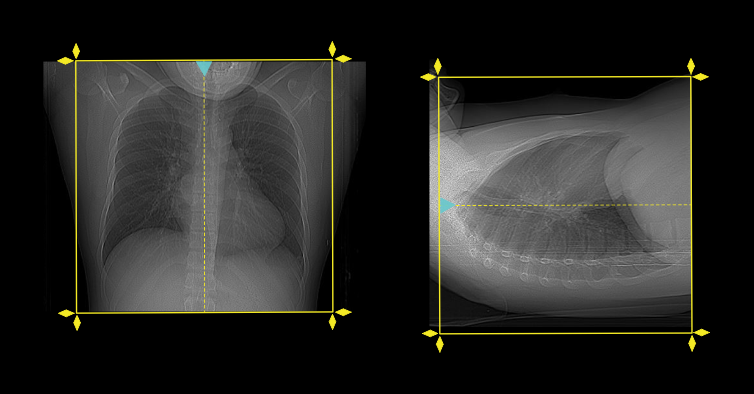
- Plan the scan starting point at the lower neck.
Scan planning
- Plan the scanning slab to cover from the lung apex to posterior costophrenic sulci.
- Acquire the scan under suspended full inspiration.
Occasionally, scan may be acquired under free breathing for both adults and children if they are unable to hold their breath, and motion artifacts of these images are lessor if the CT scanner has wider detector width, faster gantry rotation and higher pith value.
Post-processing
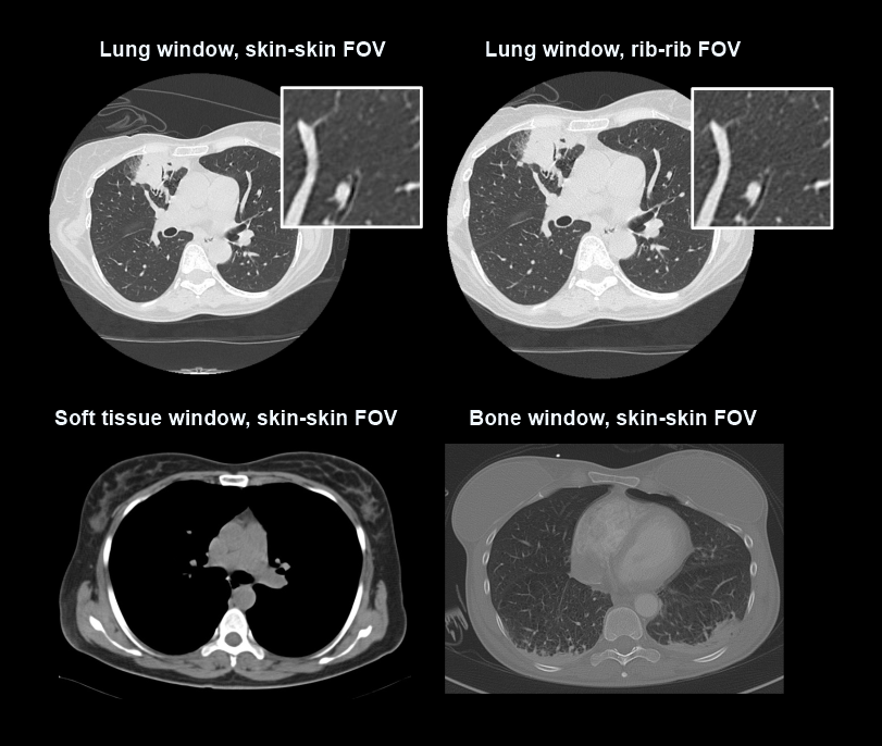
- Axial, coronal and sagittal images with <3 mm slice thickness in both lung (WW: 1500, WL: -500) and bone window (WW: 3500, WL: 350).
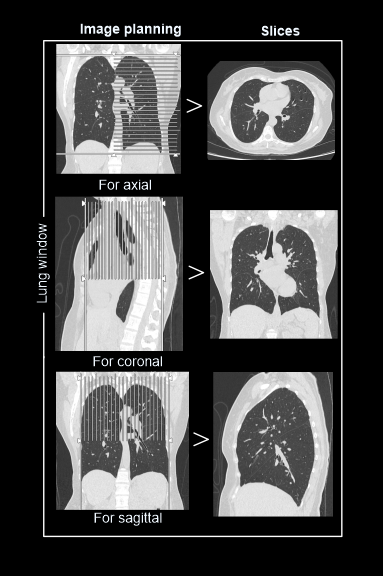
- Field of view (FOV) of lung window should be reduced to rib cage.
Explanation: to increase spatial resolution of the image, and this is specific for diffuse lung diseases.
- Axial, coronal and sagittal images with <5mm slice thickness in soft-tissue window (WW: 400, WL:40).
- FOV of soft-tissue and bone windows should include skin margins.
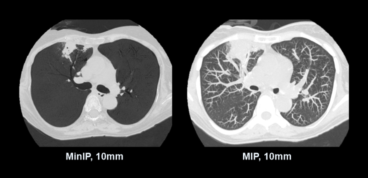
- Overlapping maximum intensity projection (MIP) images with 5-10mm slab thickness, in transverse plane.
Explanation: helps to detect lung nodules.
- Minimum intensity projection (MinIP) images with <10mm slab thickness, in transverse plane.
Explanation: helps to assess emphysema and air trapping.
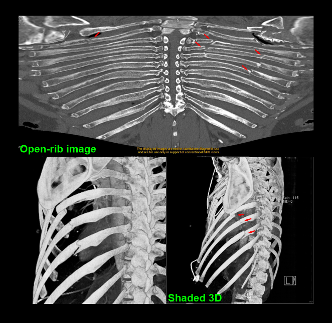
- Additionally, shaded 3D and open-rib images to visualize conditions associated with the thoracic spine and ribs.
Reference
- Mannudeep K. S. Kalra, MD, Chair, Jessica Kurian MD, & Satinder Singh, MD, FSABI. (2023). ACR–SABI–SPR–STR Practice parameter for the performance of thoracic computed tomography (CT). Retrieved from www.gravitas.acr.org.
- Bhalla, A. S., Das, A., Naranje, P., Irodi, A., Raj, V., & Goyal, A. (2019). Imaging protocols for CT chest: A recommendation.The Indian journal of radiology & imaging, 29(3), 236–246. https://doi.org/10.4103/ijri.IJRI_34_19
- Iezzi, R., Larici, A. R., Franchi, P., Marano, R., Magarelli, N., Posa, A., Merlino, B., Manfredi,R., & Colosimo, C. (2017). Tailoring protocols for chest CT applications: when and how?.Diagnostic and interventional radiology (Ankara, Turkey), 23(6), 420–427. https://doi.org/10.5152/dir.2017.16615
- Kubo T, Ohno Y, Nishino M, Lin PJ, Gautam S, Kauczor HU, Hatabu H; iLEAD study group. Low dose chest CT protocol (50 mAs) as a routine protocol for comprehensive assessment of intra thoracic abnormality.Eur J Radiol Open. 2016 Apr 27;3:86-94. doi: 10.1016/j.ejro.2016.04.001. PMID: 27957519; PMCID: PMC5144113.
- Mussmann, B., Skov, P. M., Lorentzen, M. H., Skjøt-Arkil, H., Graumann, O., Andersen, M. B., &Jensen, J. (2023). Ultra-low-dose emergency chest computed tomography protocols in three vendors: A technical note.Acta radiologica open, 12(3), 20584601231183900. https://doi.org/10.1177/20584601231183900
- Romanyukha, A., Nzitunga, P. S., & Dolcet, A. (2022, April 28). CT patient positioning plays key role in radiation dose reduction.www.auntminnie.com.