CT Head (Non-Contrast)
Last updated January 25, 2026
Similar expressions
CT head/ brain CT/ NCCT brain/ brain non-contrast enhanced
Introduction
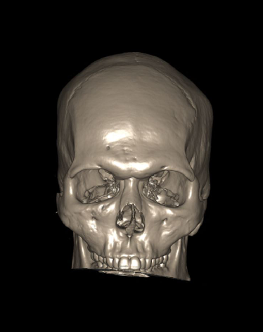
CT Brain scan is one of the routine examinations in computed tomography (CT). It is mainly referred for the diagnosis of hemorrhages, trauma, stroke and cerebral tumors.
Patient preparation
- Remove hair clips, ear rings or any removable metal in the exposing area.
- Check contraindications for radiation exposure.
- Explain the procedure clearly.
- Ask the patient to be steady during the acquisition.
Patient positioning
- Position in supine and head first on the CT couch.
- Position the head in the head rest.
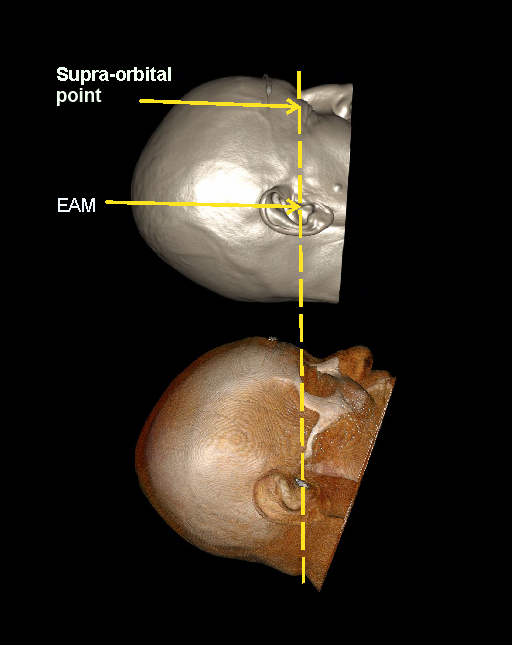
- Tilt patient’s head such that the line joining supra-orbital point and external auditory meatus (EAM) perpendicular to the floor, and ask to close the eyes.
Explanation: This method reduces eye lens dose [1 and 4].
- Immobilize the head using a strap.
- Center the scanning area in the scanner iso-center [6].
Explanation: this reduces overall radiation exposure and increases image quality.
Scan planning
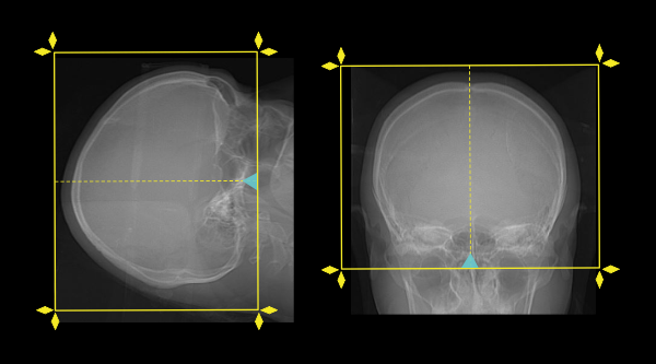
- Plan the scan starting point (laser indicator) at the upper lip of the patient.
- Plan the scan range from the skull vertex to skull base.
Post-processing
- 5 mm axial, coronal and sagittal images in brain window (WW80, WL40) [7].
- 1-2mm axial, coronal or sagittal images of the skull in bone window (WW3500, WL350) to show fractures, dislocations or bony abnormalities.
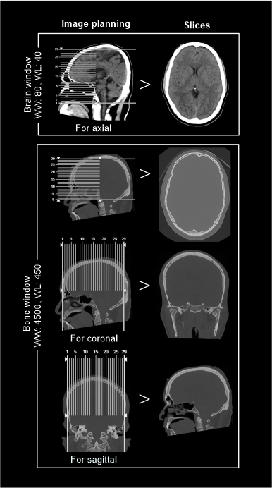
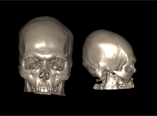
- 3d images to show fractures, dislocations or abnormalities.
Reference
- Mosher EG, Butman JA, Folio LR, Biassou NM, Lee C. Lens Dose Reduction by Patient Posture Modification During Neck CT.AJR Am J Roentgenol. 2018 May;210(5):1111-1117. doi: 10.2214/AJR.17.18261. Epub 2018 Mar 16. PMID: 29547058; PMCID: PMC8666130
- Maetani, K., Namiki, J., Matsumoto, S., Matsunami, K., Narumi, A., Tsuneyoshi, T., & Kishikawa, M. (2016). Routine Head Computed Tomography for Patients in the Emergency Room with Trauma Requires Both Thick- and Thin-Slice Images. Emergency medicine international, 2016, 5781790.
- Liebmann M, Lüllau T, Kluge A, Poppe B, von Boetticher H. Patient radiation protection covers for head CT scans - a clinical evaluation of their effectiveness. Rofo. 2014 Nov;186(11):1022-7. doi: 10.1055/s-0034-1366279. Epub 2014 Apr 1. PMID: 24691839.
- Tarkiainen, J., Nadhum, M., Heikkilä, A., Rinta-Kiikka, I., & Joutsen, A. (2023). Radiation dose of the eye lens in CT examinations of the brain in clinical practice-the effect of radiographer training to optimise gantry tilt and scan length. Radiation protection dosimetry, 199(5), 391–398.
- Booij, R., Budde, R. P. J., Dijkshoorn, M. L., & van Straten, M. (2019). Accuracy of automated patient positioning in CT using a 3D camera for body contour detection. European radiology, 29(4), 2079–2088.
- Romanyukha, A., Nzitunga, P. S., & Dolcet, A. (2022, April 28). CT patient positioning plays key role in radiation dose reduction. www.auntminnie.com.
- Wintermark, MD, M., Blumfield, E., & E. Jordan, J. (2022). ACR–ASNR–SPR Practice parameter for the performance of computed tomography (CT) in the evaluation and classification of traumatic brain injury.