CT Scan hip joint
Last updated June 03, 2026
By Radiohelp Staff
Similar expressions
CT hip joint/ NCCT hip/ CT both hips
Introduction
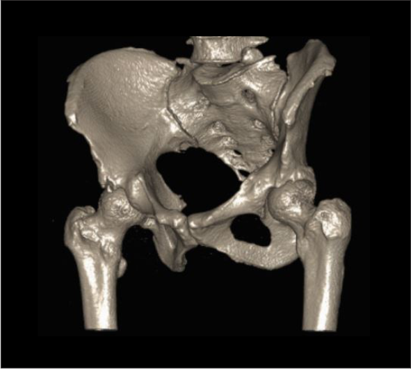
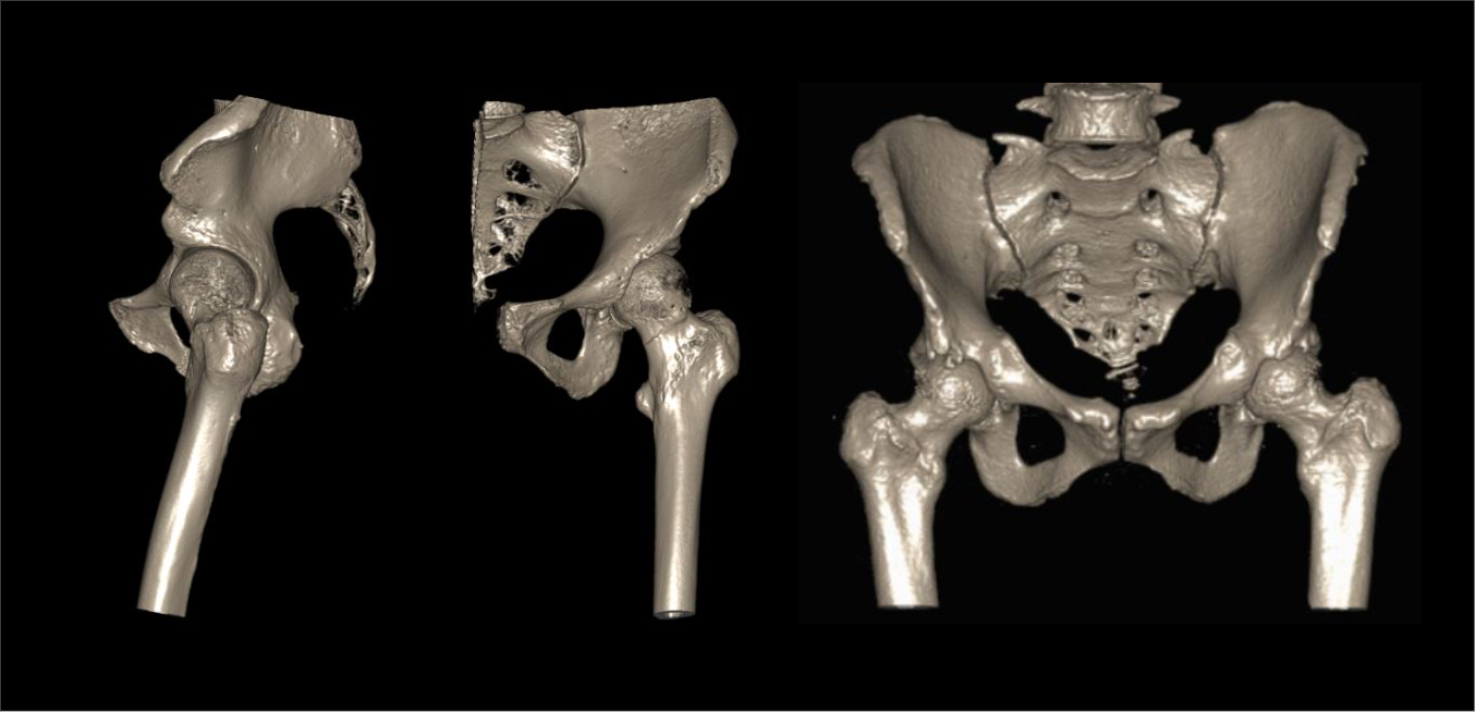
CT hip joint or CT both hips protocol is used to assess bony structure of hip joint, including fractures, neoplasms, congenital abnormalities and implants. Specially, scan has the ability to generate 3D reconstruction images that helps in surgery planning.
Patient preparation
- Assess patient’s suitability to expose radiation.
- Remove removable metallic objects in the interested region.
- Ask not to move during the scan.
Patient positioning
- Position in feet-first supine.
- Place both legs flat on the table.
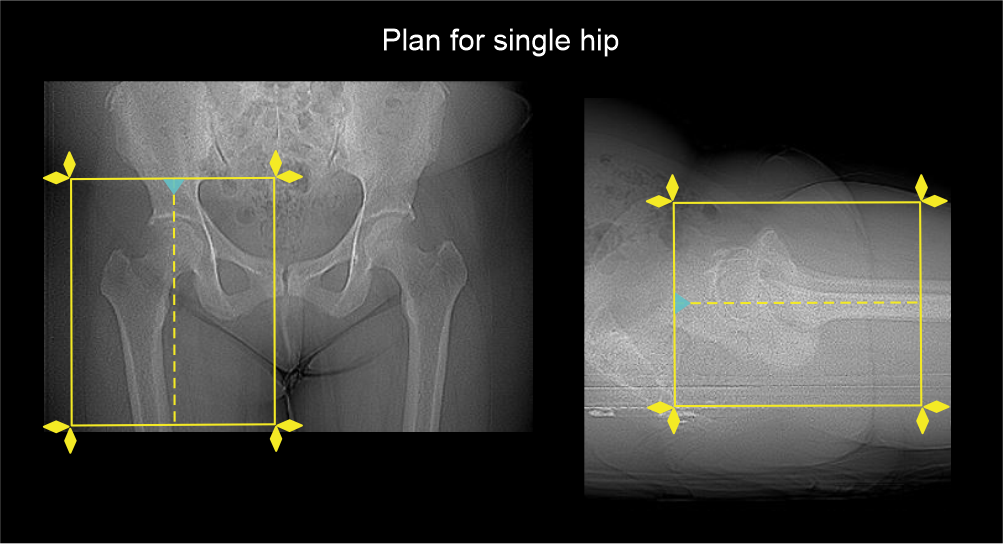
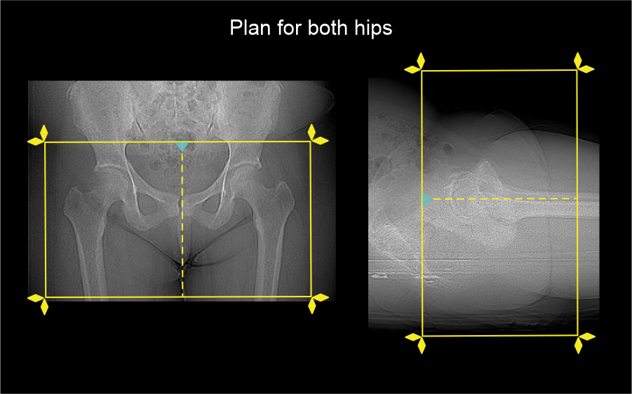
- Position pelvic region in the iso-center for both hip joint scan or affected hip joint closer to iso-center for single hip scan.
Explanation: this reduces radiation dose to patient and improves image quality.
- Rotate patient’s leg or legs internally if possible.
Explanation: this makes femoral neck parallel.
- Place a sand bag on lower limbs to immobilize.
Scan planning
- Plan the scan slab to cover from anterior inferior iliac spine (AIIS) to proximal 1/3 of femur.
- Reduce field of view (FOV) adequately to include the affected hip joint.
Explanation: this increases geometric resolution of images.
Post-processing
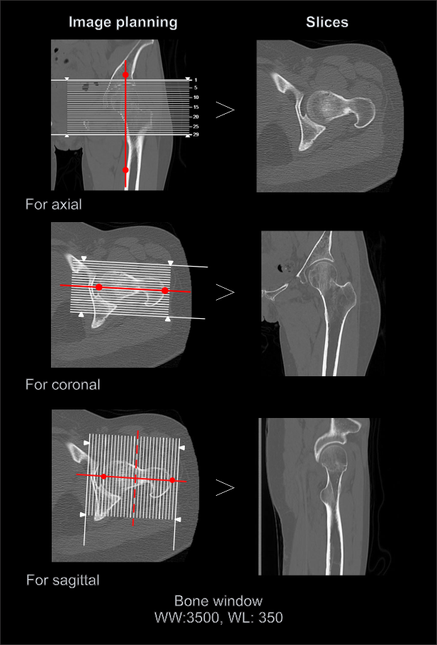
- Axial, sagittal and coronal images in both bone (WW: 3500, WL: 350) and soft tissue window (WW:500, WL:50) with ≤ 2mm slice thickness.
- 3D images to demonstrate pathologies clearly.
Reference
- Curley, A. J., Ruh, E. R., Shah, A., Disantis, A. E., Krivoniak, A., Mauro, C. S., & McClincy, M. P. (2022). A systematic approach to CT evaluation of non-arthritic hip pain. EFORT open reviews , 7(9), 653–662. https://doi.org/10.1530/EOR-22-0051
- Blum A, Meyer JB, Raymond A, Louis M, Bakour O, Kechidi R, Chanson A, Gondim-Teixeira P. CT of hip prosthesis: New techniques and new paradigms. Diagn Interv Imaging. 2016 Jul-Aug;97(7-8):725-33. doi: 10.1016/j.diii.2016.07.002. Epub 2016 Jul 20. PMID: 27451263.
- Sauser DD, Billimoria PE, Rouse GA, Mudge K. CT evaluation of hip trauma. AJR Am J Roentgenol. 1980 Aug;135(2):269-74. doi: 10.2214/ajr.135.2.269. PMID: 6773325
- Gatt, T., Cutajar, D., Borg, L., & Giordmaina, R. (2021). The Necessity of CT Hip Scans in the Investigation of Occult Hip Fractures and Their Effect on Patient Management. Advances in orthopedics, 2021, 8118147. https://doi.org/10.1155/2021/8118147
- Qu, H., & Bian, L. (2024). Comparison of CT and MRI in diagnosing occult hip fracture: a systematic review and meta-analysis. American journal of translational research, 16(7), 2745–2755. https://doi.org/10.62347/NUBB1946
- Eggenberger, E., Hildebrand, G., Vang, S., Ly, A., & Ward, C. (2019). Use of CT Vs. MRI for Diagnosis of Hip or Pelvic Fractures in Elderly Patients After Low Energy Trauma. The Iowa orthopaedic journal, 39(1), 179–183.
- Boutin, R. D., Bamrungchart, S., Bateni, C. P., Beavers, D. P., Beavers, K. M., Meehan, J. P., & Lenchik, L. (2017). CT of Patients With Hip Fracture: Muscle Size and Attenuation Help Predict Mortality. AJR. American journal of roentgenology, 208(6), W208–W215. https://doi.org/10.2214/AJR.16.17226