CT Coronary Calcium
Last updated June 27, 2026
By Radiohelp Staff
Similar expressions
CT calcium scoring, CT Ca score, CT coronary calcium scoring, CCS
Introduction
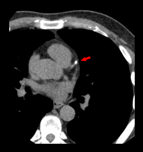
Coronary Calcium Scoring, which is a non-contrast study, is helpful to quantify coronary artery calcium deposits. It’s used for the risk stratification and therapeutic decision making in patients who are in risk of atherosclerotic cardio vascular risk. Moreover, in Transcatheter Aortic Valve Replacement (TAVR), calcium scoring is used to analyze the score of the aortic valve.
Patient preparation
- Explain the procedure clearly and kindly.
- Check the suitability to expose radiation.
- Ask to remove any metals related to the interested region.
- Ask to refrain from taking caffein for at least 6 hours prior.
Explanation: Caffein increases patient’s heart rate, making the heart to move fast during image acquisition, which causes motion artifacts in coronary arteries.
- Practice breathing technique (arrested full inspiration) properly.
Explanation: poor breadth hold can cause motion artifacts due to diaphragm movement alongwith the heart.
- Place the ECG leads and they need to be outside the scanning area.
Explanation: ECG leads has metallic components, which provide metallic artifacts. Although, ECG gating is essential in CT Ca scoring to reduce motion artifacts of the beating heart. Reach our CT coronary angiogram article for more information.
Heart rate reducing drugs such as Beta blockersaren’t mostly used because its action for this scan is not significant.
Patient positioning
- Position the patient in supine and feet first on the imaging couch.
- Raise both hands above the head and place a positioning aid under them.
Explanation: hands beside the trunk give streak artifacts and increase radiation exposure.
- Center the scanning area in the scanner iso-center [6].
Explanation: this reduces overall radiation exposure and increases image quality.
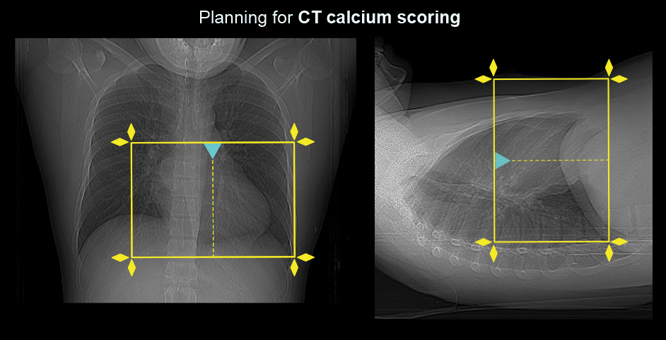
- Place the scout-initiation point at the level of supra sternal notch.
Scan planning
- Plan the scanning slab to cover from a point just below the carina and extending inferior to the left ventricle (volume scan).
- Perform heart-rate analysis during full inspiration.
Explanation: image acquisition happens when the patient is under a full inspiration, where the mean heart rate reduces approximately by 4 beats. This assessment is essential to select the appropriate cardiac window, and scanner selects the phase automatically. Reach CT coronary angiogram article for illustrations.
Scan uses prospective gating for imaging that aquieres a narrow cardiac window (mostly 75%), resulting a very low radiation dose compaired to CT coronary angigraphy. Please read our CT coronary angiography article if you need more information regarding cardiac-gating, cardiac-windo, cardiac-cycle and padding.
Post-processing
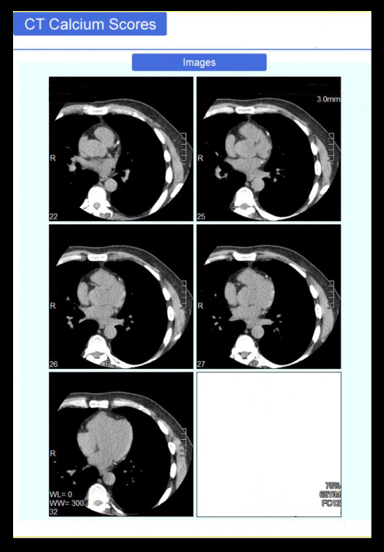
- Axial images in cardiac window (WW: 1000, WL: 400) with 3mm slice thickness.
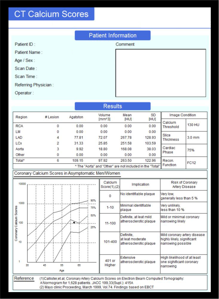
- Calcium score analysis report.
Reference
- Neves, P. O., Andrade, J., & Monção, H. (2017). Coronary artery calcium score: current status.Radiologia brasileira, 50(3), 182–189. https://doi.org/10.1590/0100-3984.2015.0235
- Koopman, M. Y., Reijnders, J. J. W., Willemsen, R. T. A., van Bruggen, R., Doggen, C. J. M., Kietselaer, B., Oude Wolcherink, M. J., van Ooijen, P. M. A., Gratama, J. W. C., Braam, R., Oudkerk,M., van der Harst, P., Dinant, G. J., & Vliegenthart, R. (2022). Coronary calcium scoring as first-line test to detect and exclude coronary artery disease in patients presenting to the general practitioner with stable chest pain: protocol of the cluster-randomised CONCRETE trial.BMJ open, 12(4), e055123. https://doi.org/10.1136/bmjopen-2021-055123
- Divakaran, S., Cheezum, M. K., Hulten, E. A., Bittencourt, M. S., Silverman, M. G., Nasir, K., & Blankstein, R. (2015). Use of cardiac CT and calcium scoring for detecting coronary plaque: implications on prognosis and patient management.The British journal of radiology, 88(1046), 20140594. https://doi.org/10.1259/bjr.20140594
- Larissa Braga Casaburi, MD, MPH, MHA, Co-Chair, Andrew L. Rivard, MD, Co-Chair, & Dhiraj Baruah, MD. (2022). ACR–NASCI–SPR Practice parameter for the performance of quantification of cardiovascular computed tomography (CT) and magnetic resonance imaging (MRI).Retrieved from ww.gravitas.acr.org.
- Klaus Hagspiel, MD, Chair, Lucia Flors Blasco, MD, PhD, & Cristina Fuss, MD. (2021). ACR–NASCI–SPR Practice parameter for the performance and interpretation of cardiac computed tomography (CT).Retrieved from www.gravitas.acr.org.
- Romanyukha, A., Nzitunga, P. S., & Dolcet, A. (2022, April 28). CT patient positioning plays key role in radiation dose reduction.www.auntminnie.com.
- Matsuura N, Horiguchi J, Yamamoto H, Hirai N, Tonda T, Kohno N, Ito K. Optimal cardiac phase for coronary artery calcium scoring on single-source 64-MDCT scanner: least interscan variability and least motion artifacts.AJR Am J Roentgenol. 2008 Jun;190(6):1561-8. doi: 10.2214/AJR.07.3120. PMID: 18492907.