CT Head venogram
Last updated April 10, 2026
Similar expressions
CT head venogram/ CT cerebral venogram/ CTV head/ CT venography brain
Introduction
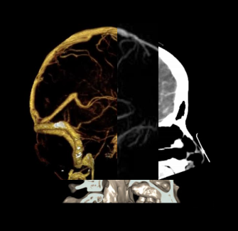
CT head venogram is a contrast study that helps to visualize cerebral venous system. Scan is useful in the diagnosis of cerebral venous thrombosis and anatomical assessments.
Patient preparation
- Explain the procedure clearly and kindly.
- Prepare the patient for contrast media administration before the procedure.
- Place a ‘green cannula’ (18G) in a stable vein with the ability of administering intravenous (IV) contrast at a higher flow rate.
Patient positioning
- Position the patient in head first supine and head on the head rest.
- Secure the head with immobilizing straps.
- Place both hands beside the body.
- Center the scanning area in the scanner iso-center.
Explanation: this reduces overall radiation exposure and increases image quality [1].
- Hand, which is used to infuse contrast media, is placed straight to facilitate contrast flow.
Scan planning
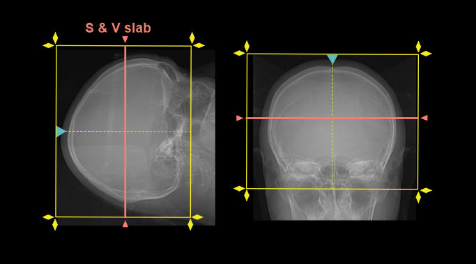
- Plan the scan slab to cover from a point just below skull base to skull vertex.
- Plan the scan and view (S & V) slab in the mid part of the head.
Explanation: Superior sagittal sinus need to be visualized in the axial cross section.
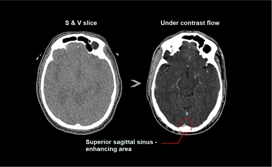
- Manual start is used to initiate the scan because it is quite challenging to find cerebral venous system in S & V slice (non-contrast), and also to place a small region of interest (ROI).
Explanation: pay attention to the contrast flow in S & V slice, start the scan when you see a slight contrast enhancement in the superior sagittal sinus.
- Scan direction is planned to initiate from the skull vertex to skull base.
Explanation: it reduces the risk of missing the whole cerebral venous phase.
- Plan 10 seconds from contrast media injection to S & V initiation.
- Plan minimum or zero delay time to start the helical venogram.
Contrast media administration
- 50ml to 60ml contrast volume can be used with a 4.5 ml/s flow rate (patient body weight 50-60kg).
Explanation: higher flow rates are used in venography to increase venous visualization.
Acquisition of both arterial and venous cerebral systems using a single contrast injection
- An extra scan slab, which is for venous acquisition, is added to the Cerebral angiography scan.
- Plan venous-scan slab to cover from a point just below skull base to skull vertex.
- Plan venous-scan direction from skull base to skull vertex.
Explanation: remember, arterial scan direction should be vertex to skull base.
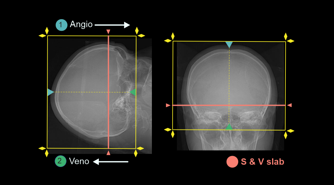
- Set a minimum or zero-time delay for venous scan.
Explanation: This minimizes the risk of missing the venous phase.
- Scan initiation is same as Cerebral angiography.
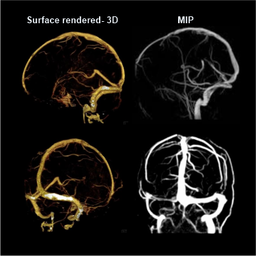
Post-processing
- Multiple images in shaded volume (3D) and maximum intensity projection (MIP) with a viewing angle of 360 degree.
- Axial, coronal and sagittal images in soft-tissue window (WW400, WL40) with 2-3mm slice thickness (refer Brain contrast article for images).
- Additionally, multiplanar reformation (MPR) images in bone window (WW4500, WL450) with 1-2mm slice thickness.
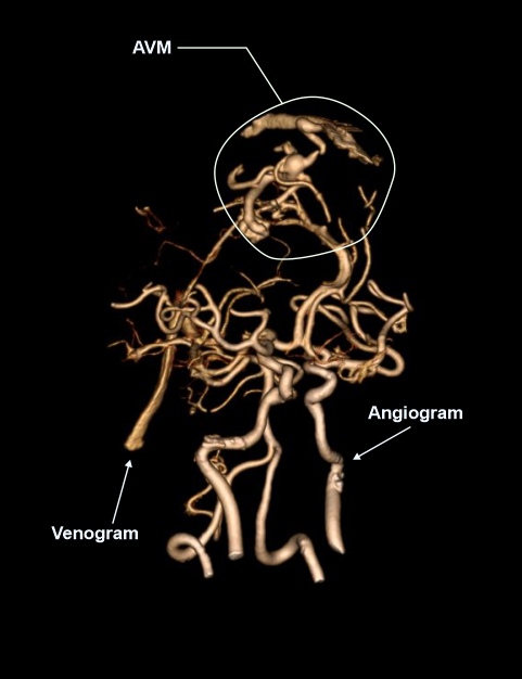
Advance post processing software can be used to separate venous system from arterial system. However, including both phases in the same image is also helpful to diagnose arteriovenous malformations (AVM).
Reference
- Romanyukha, A., Nzitunga, P. S., & Dolcet, A. (2022, April 28). CT patient positioning plays key role in radiation dose reduction.
- Biswas S, Chandran A, Roughley S, Bhojak M, Das K. Cerebral CT Venography Using a 320-MDCT Scanner with a Time-Density Curve Technique and Low Volume of Contrast Agent: Comparison with Fixed Time-Delay Technique.AJR Am J Roentgenol. 2015 Dec;205(6):1269-75. doi: 10.2214/AJR.14.14200. PMID: 26587933.
- Wetzel, S. G., Kirsch, E., Stock, K. W., Kolbe, M., Kaim, A., & Radue, E. W. (1999). Cerebral veins: comparative study of CT venography with intraarterial digital subtraction angiography. AJNR. American journal of neuroradiology, 20(2), 249–255.
- Blackham, MD, Chair, K., Jordan, MD, FACR, J., & Corey, MD, FACR, A. (Eds.). (2020). ACR–ASNR–SPR Practice parameter for the performance of computed tomography (CT) of the head.
- Seo H, Choi DS, Shin HS, Cho JM, Koh EH, Son S. Bone subtraction 3D CT venography for the evaluation of cerebral veins and venous sinuses: imaging techniques, normal variations, and pathologic findings. AJR Am J Roentgenol. 2014 Feb;202(2): W169-75. doi: 10.2214/AJR.13.10985. PMID: 24450700.