Adrenal CT scan Protocol
Last updated July 01, 2026
By Radiohelp Staff
Similar expressions
CT adrenals/ CT adrenal scan
Introduction
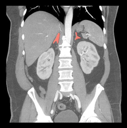
Adrenal CT protocol helps to characterize masses in the adrenal glands. This study has multiple pre and post contrast phases that are used to identify contrast washout of masses.
Patient preparation
- Explain the procedure clearly and kindly.
- Check the suitability to administer intra-venous (IV) iodinated contrast.
- Place an Intra venous (IV) cannula in a stable vein for contrast injection – pink-20G cannula.
- Instruct the patient to breath in and hold during the scan.
Explanation: avoids motion un-sharpness of liver, pancreas and kidneys.
- Explain the burning sensation during contrast injection.
- Check the ability to expose radiation.
- Ask the patient to remove metals related to the interested region.
- Use gastro-intestinal contrast media if needed – refer multiphase contrast abdomen article for their usage.
Patient positioning
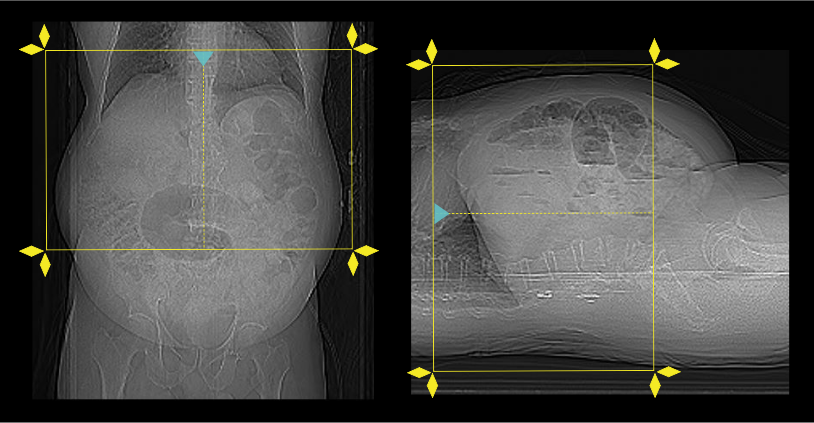
- Position the patient in feet first and supine position.
- Center the scanning area in the scanner iso-center [6].
Explanation: this reduces overall radiation exposure and increases image quality.
- Place both hands above the head.
Explanation: this reduces streak artifacts from the hands, also it avoids unnecessary radiation exposure to the hands.
- Keep the arm with the IV cannula strait.
Explanation: to facilitate contrast flow.
- Plan the scout starting point at the nipple level.
Scan planning
Usually, there are three steps in the scan: pre-contrast, venous and delay.
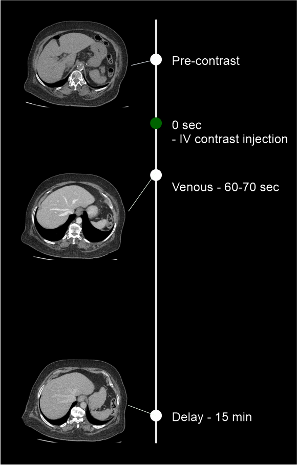
- Plan all three steps to cover from the dome of the diaphragm to iliac crest.
- Set phase initiation using bolus timing method – refer multiphase contrast abdomen scan for more details.
Intravenous (IV) contrast administration
- Generally, a volume of 75 to 80 ml of iodinated-contrast can be injected at a flow rate of 3-3.5ml/s – this is for a patient with a body weight of 75 to 80 kg.
- Inject 50ml of saline flush following the contrast injection.
Explanation: flushes remaining contrast in the veins of the injected hand, and maintainscontrast flow for a longer time.
- Conduct all phases under arrested inspiration.
Post-processing
A qualified Radiologist must calculate the Hounsfield unit (HU) values of adrenal lesions for all three phases to identify the contrast washout.
- Multiplanar reconstruction (MPR) images for all phases in soft-tissue window (WW:500, WL:50) with slice thickness 1.5-3mm.
- Axial reconstruction in bone window (WW: 3500, WL: 350) with ≤ 3mm slice thickness.
Refer non-contrast abdomen and pelvis scan for images.
Reference
- Olga R. Brook, MD, Chair, Jessica Kurian MD, Alec Megibow, MD, MPH, FACR, & Michael Furman,MD. (2021). ACR–SABI–SAR–SPR practice parameter for the performance of computed tomography (CT) of the abdomen and computed tomography (CT) of the pelvis.Retrieved from www.gravitas.acr.org.
- Schloetelburg, W., Ebert, I., Petritsch, B., Weng, A. M., Dischinger, U., Kircher, S., Buck, A.K., Bley, T. A., Deutschbein, T., & Fassnacht, M. (2021). Adrenal wash-out CT: moderate diagnostic value in distinguishing benign from malignant adrenal masses.European journal of endocrinology, 186(2), 183–193. https://doi.org/10.1530/EJE-21-0650
- James T. Lee, P. L., Michael Corwin, Co-chair, Elaine Caoili, Co-Chair, Eric Remer, & Dan Glazer. (2022, 03). CT adrenal mass protocols v1.0, Society of Abdominal Radiology Disease Focused Panel on Adrenal Neoplasm.Retrieved from www.abdominalradiology.org.
- Nanda Krak, & Robin Smithuis. Characterization of Adrenal lesions. Retrieved from www.radiologyassistant.nl.
- Panda, A., Das, C. J., Dhamija, E., Kumar, R., & Gupta, A. K. (2015). Adrenal imaging (Part 1): Imaging techniques and primary cortical lesions.Indian journal of endocrinology and metabolism, 19(1), 8–15. https://doi.org/10.4103/2230-8210.146858
- Romanyukha, A., Nzitunga, P. S., & Dolcet, A. (2022, April 28). CT patient positioning plays key role in radiation dose reduction.www.auntminnie.com.