CT Shoulder Joint Scan
Last updated June 03, 2026
By Radiohelp Staff
Similar expressions
CT shoulder joint/ NCCT shoulder/ CT SJ
Introduction
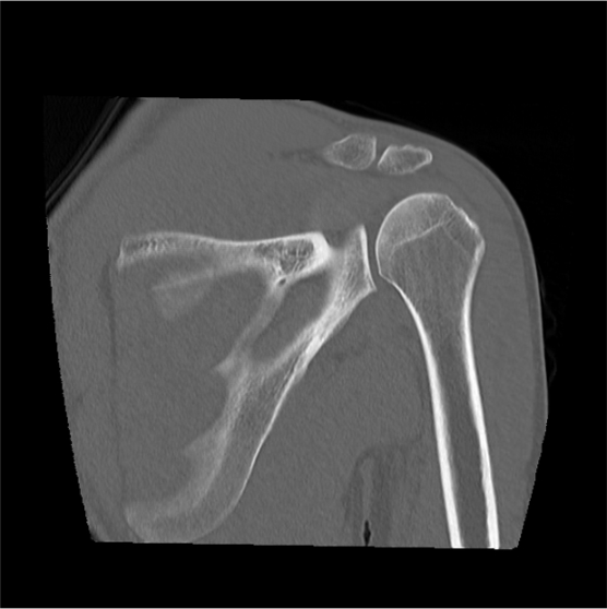
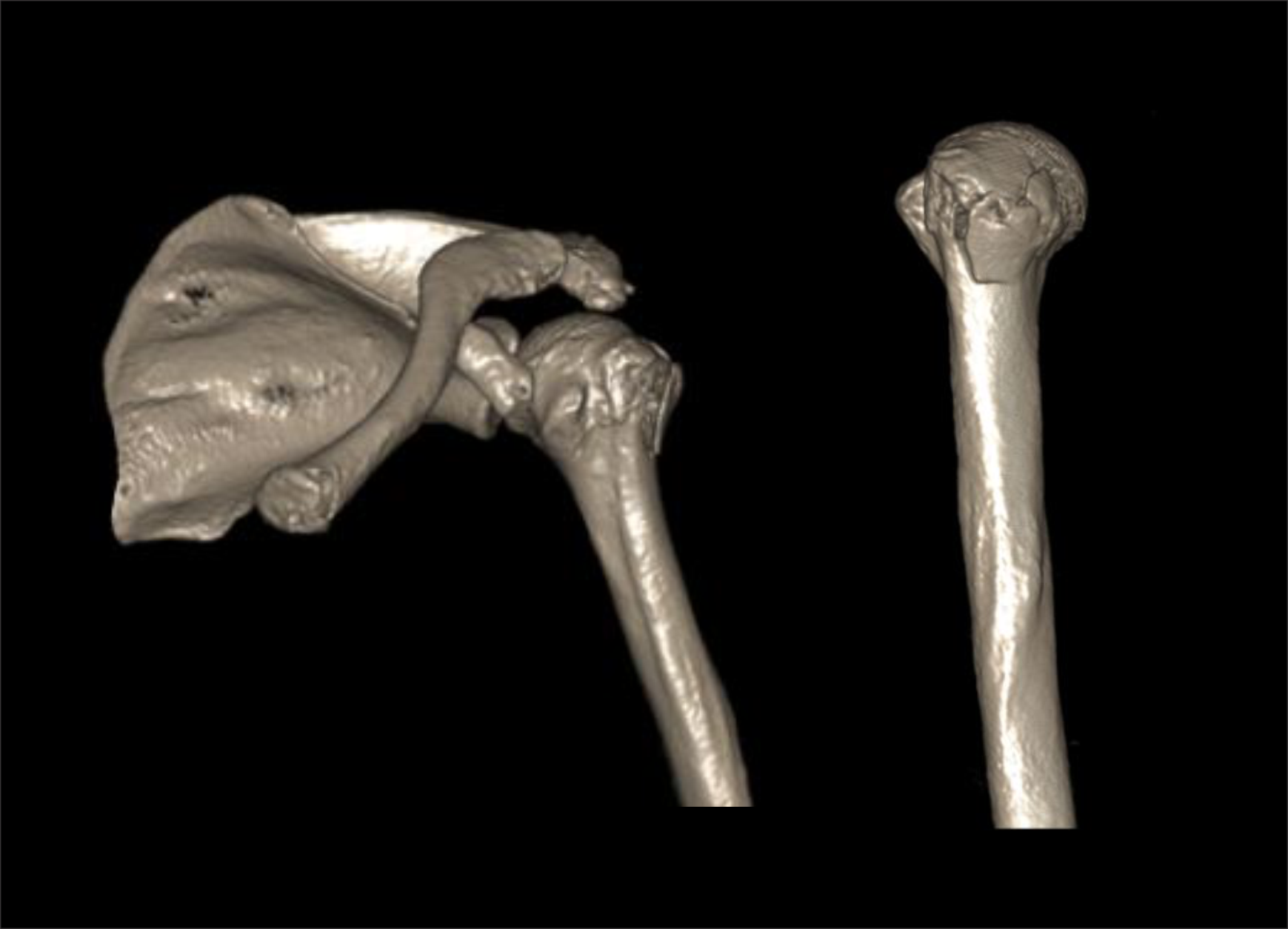
NCCT shoulder scan is used to evaluate fractures, implant complications and neoplasm. Its ability to view structures in three dimension (3D) is remarkable for surgical planning.
Patient preparation
- Explain the procedure kindly and clearly.
- Remove radio opaque items related to the shoulder area including necklaces and under garments.
Patient positioning
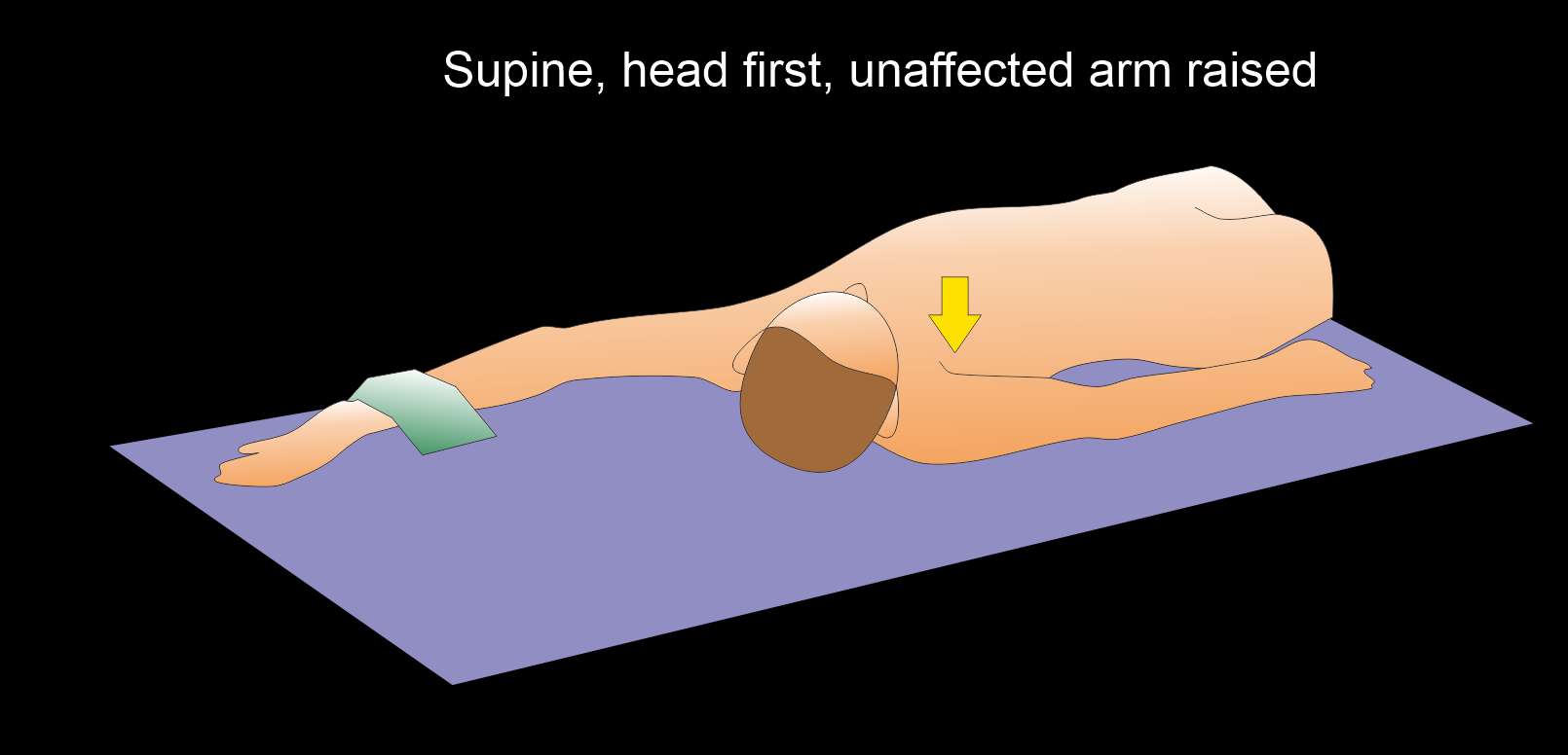
- Position in feet first, supine and move the affected shoulder slightly towards the iso-center.
Explanation: shoulder in the iso-center reduces radiation dose and increases image quality.
- Raise the unaffected arm above the head.
Explanation: this reduces radiation dose and streak artifacts.
- Place the affected arm beside the body and rotate externally (thumb points outward or anatomical position).
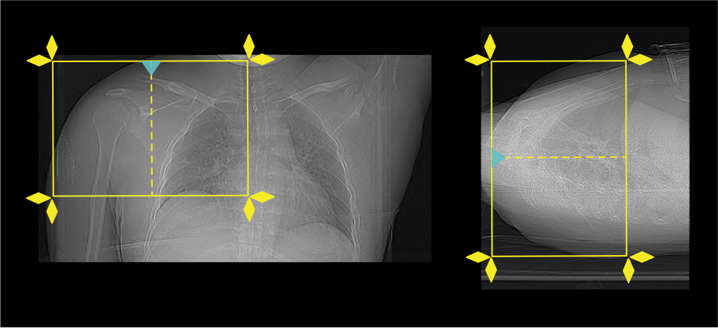
- Set the scout start point at the nipple level and scan direction into the gantry.
Scan planning
- Plan the scan slab to cover from the acromioclavicular joint (ac joint) to inferior angle of the scapula.
- Reduce the field of view (FOV) as appropriate to include scapula, clavicle and humerus.
Explanation: small FOV increases geometric resolution.
Post-processing
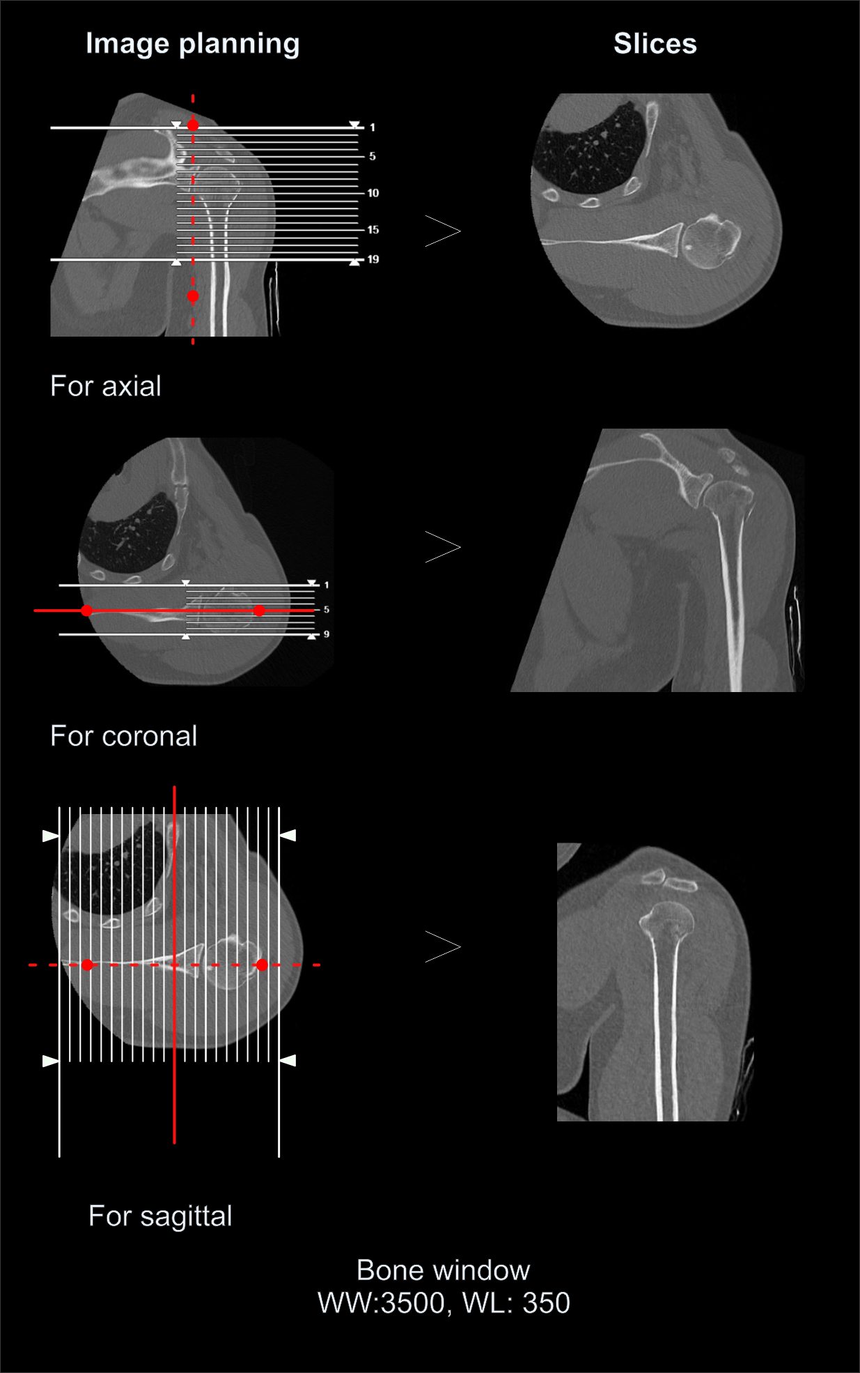
- Coronal, sagittal and axial images in both soft tissue (WW:500, WL:50) and bone (WW: 3500, WL: 350) window with ≤2mm slice thickness.
- 3d images to show pathologies clearly.
Reference
- Gregory, T., Hansen, U., Khanna, M., Mutchler, C., Urien, S., Amis, A. A., Augereau, B., & Emery, R. (2014). A CT scan protocol for the detection of radiographic loosening of the glenoid component after total shoulder arthroplasty. Acta orthopaedica, 85(1), 91–96. https://doi.org/10.3109/17453674.2013.869653
- Slocum, A. M. Y., Siu, Y. C., Ma, C. M., & Lui, T. H. (2021). The study of 2-dimensional computed tomography scans of the glenoid anatomy in relation to reverse shoulder arthroplasty in the Southern Chinese population. JSES international, 5(4), 714–721. https://doi.org/10.1016/j.jseint.2021.02.006
- Rafii M, Minkoff J. Advanced arthrography of the shoulder with CT and MR imaging. Radiol Clin North Am.1998 Jul;36(4):609-33. doi: 10.1016/s0033-8389(05)70052-x. PMID: 9673643.
- Bao MH, DeAngelis JP, Wu JS. Imaging of traumatic shoulder injuries - Understanding the surgeon's perspective.Eur J Radiol Open. 2022 Mar 2;9:100411. doi: 10.1016/j.ejro.2022.100411. PMID: 35265737; PMCID: PMC8899241.
- Xiao, M., Zhang, M., Lei, M., Lin, F., Chen, Y., Chen, J., Liu, J., & Ye, J. (2023). Diagnostic accuracy of ultra-low-dose CT compared to standard-dose CT for identification of non-displaced fractures of the shoulder, knee, ankle, and wrist. Insights into imaging, 14(1), 40. https://doi.org/10.1186/s13244-023-01389-7
- Romanyukha, A., Nzitunga, P. S., & Dolcet, A. (2022, April 28). CT patient positioning plays key role in radiation dose reduction.www.auntminnie.com.