CT Hand scan
Last updated June 03, 2026
By Radiohelp Staff
Similar expressions
CT hand/ NCCT hand
Introduction
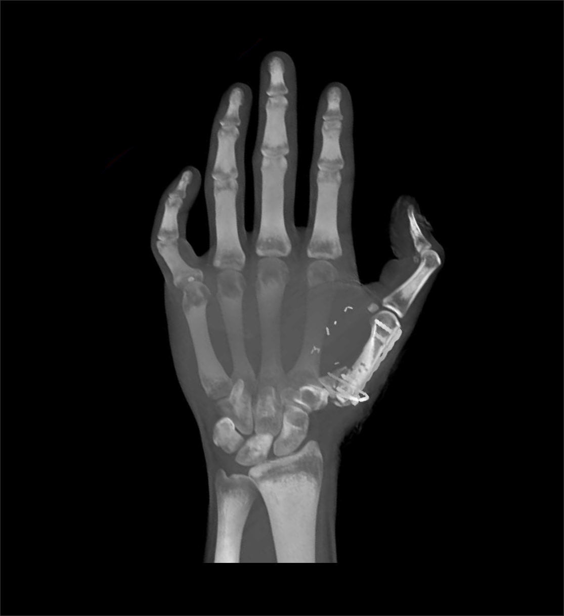
CT hand protocol is used to evaluate conditions such as fractures, implant complications and neoplasm. Its ability to view structures in three dimension (3D) is remarkable for surgical planning.
Patient preparation
- Explain the procedure kindly and clearly.
- Remove radio opaque items related to hand and wrist area.
Patient positioning
There are several positioning methods.
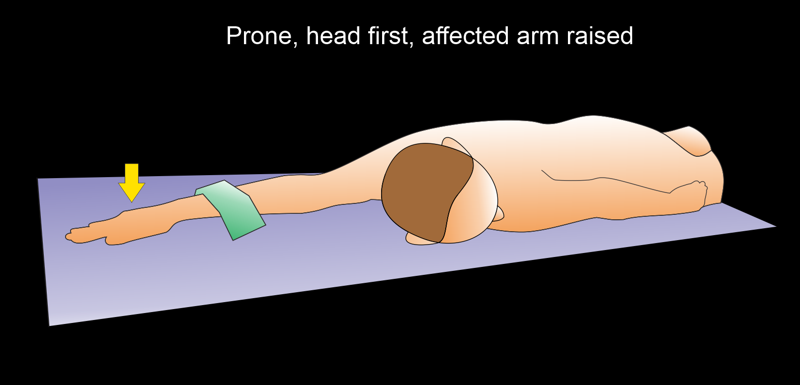
Method 1 - patient in prone, head first
- Raise the affected arm above the head and keep the unaffected arm besides the body.
- Center the scan area in the iso-center.
Explanation: hand in the iso-center helps to reduce radiation exposure and increase image quality.
- Pronate the affected hand-fingers straight and close together.
- Bend patient’s head towards the unaffected arm.
- Place a sand bag on the proximal one third of the forearm (affected arm) to immobilize.
- Ask not to move fingers or hand.
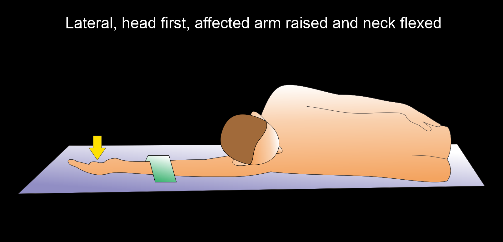
Method 2 - patient in lateral decubitus, head first
- Position the patient in lateral decubitus on the affected side.
- Raise the affected arm above the head.
- Supinate the hand-fingers straight and close together.
- Place a sand bag on the proximal one third of the affected forearm to immobilize.
- Center the scan area in the iso-center.
- Ask not to move fingers or hand.
Explanation: this method is suitable for the patients who are unable to be in prone position.
Scan planning
- Plan the scan slab to cover from the distal radioulnar joint to fingertips.
- Reduce the field of view (FOV) as appropriate to include the hand.
Explanation: reduced FOV increases geometric resolution.
Post-processing
- Coronal, sagittal and axial images in both soft tissue (WW:500, WL:50) and bone (WW: 3500, WL: 350) window with ≤2mm slice thickness.
- 3d images to show pathologies clearly.
Reference
- Münn, F., Laun, R. A., Asmus, A., Bülow, R., Bakir, S., Haralambiev, L., Eisenschenk, A., & Kim, S. (2020). Detection of fractures of hand and forearm in whole-body CT for suspected polytrauma in intubated patients.BMC musculoskeletal disorders, 21(1), 49. https://doi.org/10.1186/s12891-020-3068-0
- Miwa S, Otsuka T. Practical use of imaging technique for management of bone and soft tissue tumors.J Orthop Sci. 2017 May;22(3):391-400. doi: 10.1016/j.jos.2017.01.006. Epub 2017 Feb 1. PMID: 28161235.
- You, J. S., Chung, S. P., Chung, H. S., Park, I. C., Lee, H. S., & Kim, S. H. (2007). The usefulness of CT for patients with carpal bone fractures in the emergency department. Emergency medicine journal : EMJ, 24(4), 248–250. https://doi.org/10.1136/emj.2006.040238
- Jakubietz MG, Mages L, Zahn RK, Kenn W, Jakubietz RG, Meffert RH. The role of CT scan in postoperative evaluation of distal radius fractures: Retrospective analysis in regard to complications and revision rates.J Orthop Sci. 2017 May;22(3):434-437. doi: 10.1016/j.jos.2016.12.024. Epub 2017 Jan 20. PMID: 28117126.5).
- Cavalcanti Kußmaul, A., Kuehlein, T., Langer, M. F., Ayache, A., Löw, S., & Unglaub, F. (2024). The Conservative and Operative Treatment of Carpal Fractures. Deutsches Arzteblatt international, 121(18), 594–600. https://doi.org/10.3238/arztebl.m2024.0102
- Romanyukha, A., Nzitunga, P. S., & Dolcet, A. (2022, April 28). CT patient positioning plays key role in radiation dose reduction.www.auntminnie.com.
- Xiao, M., Zhang, M., Lei, M., Lin, F., Chen, Y., Chen, J., Liu, J., & Ye, J. (2023). Diagnostic accuracy of ultra-low-dose CT compared to standard-dose CT for identification of non-displaced fractures of the shoulder, knee, ankle, and wrist. Insights into imaging, 14(1), 40. https://doi.org/10.1186/s13244-023-01389-7