CT IAM (internal auditory meatus)
Last updated May 13, 2026
Similar expressions
CT IAM/ CT IAC/ CT internal auditory meatus/ NCCT IAM/ HRCT of the temporal bone/ CT temporal bone
Introduction
CT IAM is used to assess pathologies associated with the internal auditory meatus (IAM), and scan is referred for patients with vertigo, hearing loss and tinnitus.
Patient preparation
- Remove hair clips, ear rings or any removable metal in the exposing area.
- Explain the procedure clearly.
- Ask the patient to be steady during the acquisition.
Patient positioning
- Position the patient in head first and supine on the scanner couch.
- Position the head in the head rest and use immobilizing bands.
- Tilt the head such that the line joining supra-orbital point and external auditory meatus (EAM) perpendicular to the floor, and ask to close the eyes (please find more information from our Brain non-contrast article).
Explanation: this method reduces the eye lens dose.
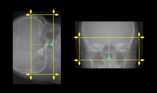
Scan planning
- Plan the scout starting point (laser indicator) at the upper lip of the patient.
- Plan the scan range to include the temporal bone that covers superiorly from the bony part of the IAM-covering the most superior mastoid air cell to mastoid tip inferiorly.
Post-processing
- Maximum slice thickness of 1mm in axial and coronal planes, reconstructed in bone window (WW:4500, WL:450) to show both canals in the same plane.
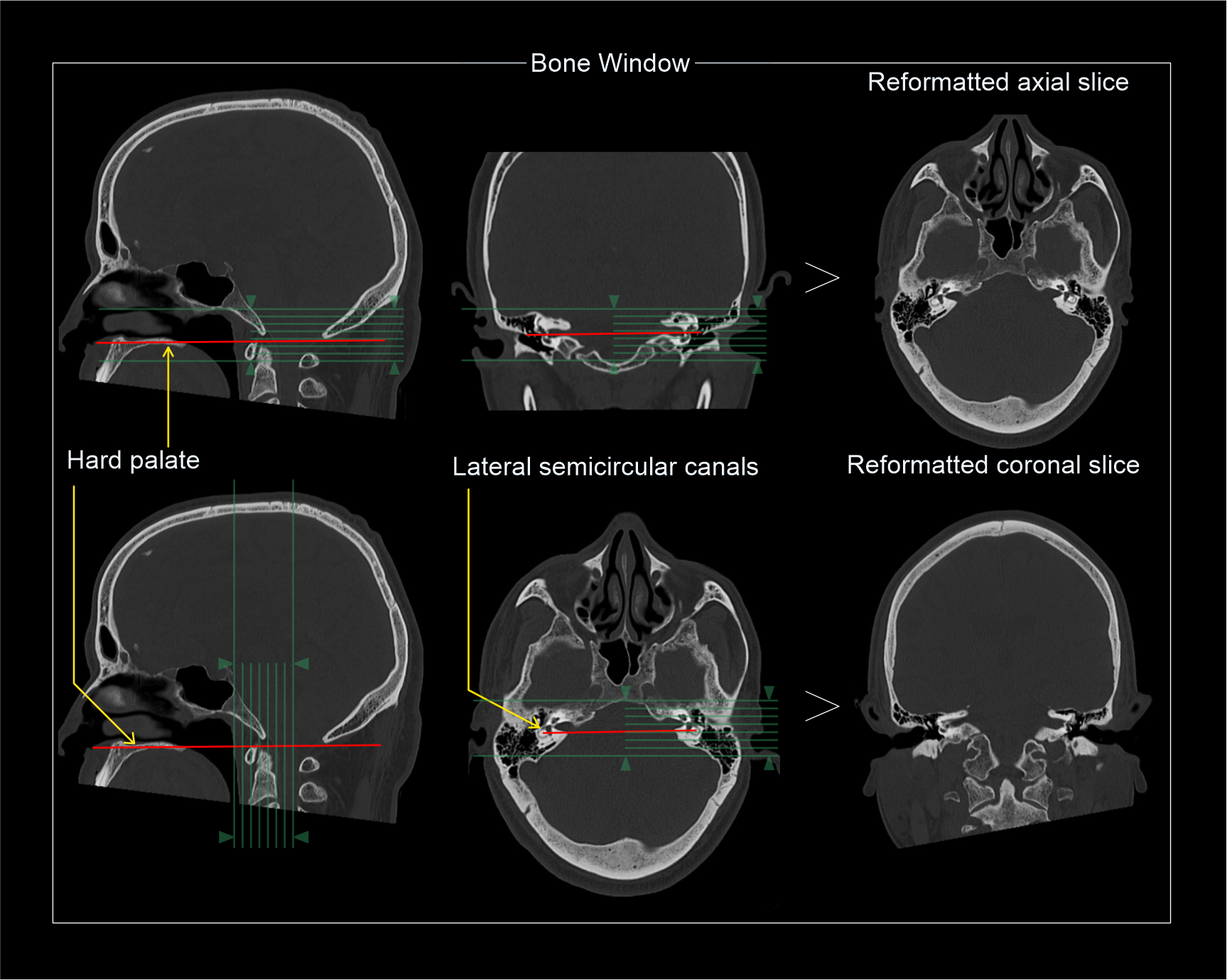
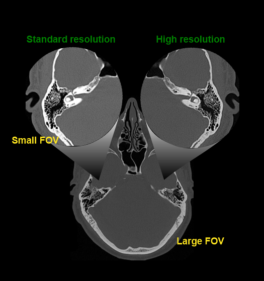
Explanation: Small-FOV reconstruction and high-resolution algorithms can be used to improve the image quality.
- Axial images of the posterior fossa in soft-tissue window (WW:400, WL:40) with 1mm slice thickness.
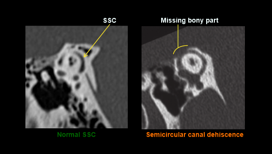
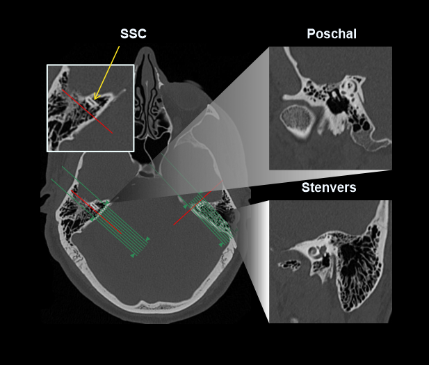
For the diagnosis of superior semicircular canal (SSC) dehiscence, which is caused due to an abnormal opening from SSC into the brain. Additional oblique images can be reformatted through SSC in short and long axis, respectively known as Poschl (Parallel to the plane of SSC) and Stenvers (Perpendicular to the plane of SCC).
Reference
- Ashley H. Aiken, MD, Chair, Paul M. Bunch, MD, & Kavita K. Erickson, MD. (2021). ACR–ASNR–SPR Practice parameter for the performance of computed tomography (CT) of the extracranial head and neck.Retrieved from www.gravitas.acr.org.
- Bi, Wenya Linda & Brewster, Ryan & Poe, Dennis & Vernick, David & Lee, Daniel &Corrales, C & Dunn, Ian. (2017). Superior semicircular canal dehiscence syndrome.Journal of neurosurgery. 127. 1-9. 10.3171/2016.9. JNS16503.
- Teszler, Chris & Daval, Mary & Altabaa, Khaled & Williams, Marc & Ayache, Denis. (2008). Computed tomography-based workup of conductive hearing loss with normal middle ear: Don't forget superior semicircular canal dehiscence.The international tinnitus journal. 14. 53-6.
- Panara K, Hoffer M. Anatomy, Head and Neck, Ear Internal Auditory Canal (Internal Auditory Meatus, Internal Acoustic Canal) [Updated 2023 Aug 28]. In: StatPearls [Internet]. Treasure Island (FL): StatPearls Publishing; 2024 Jan.
- Romanyukha, A., Nzitunga, P. S., & Dolcet, A. (2022, April 28). CT patient positioning plays key role in radiation dose reduction. www.auntminnie.com.