CT Orbits
Last updated June 27, 2026
By Radiohelp Staff
Similar expressions
NCCT orbits/ CT orbits
Introduction
Orbits CT scan is used to diagnose conditions associated with the orbital area. Scan is helpful for the visualization of tumors, trauma, orbital foreign body and vision impairment.
Patient preparation
- Remove hair clips, ear rings or any removable metal in the exposing area.
- Explain the procedure clearly.
- Ask the patient to be steady during the acquisition.
Patient positioning
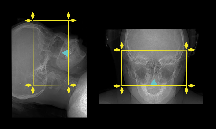
- Position the patient in head first and supine on the scanner couch.
- Position the head in the head rest and use immobilizing bands.
- Tilt the head such that the line joining supra-orbital point and external auditory meatus (EAM) perpendicular to the floor, and ask to close the eyes (please find more information from our Brain non-contrast article).
Explanation: this method reduces the eye lens dose.
- Center the scanning area in the scanner iso-center [5].
Explanation: this reduces overall radiation exposure and increases image quality.
- Ask not to move the eye balls during the scan.
Explanation: this’s to minimize the motion artifacts.
Scan planning
- Plan the scan slab to cover from hard palate up to the superior part of the frontal sinuses.
Post-processing
- Axial, sagittal and coronal Images without exceeding 3mm slice thickness, reformatted in soft-tissue (WW: 400, WL: 40) and bone window (WW: 4500, WL: 450).
Explanation: for the evaluation of foreign bodies in orbital area, reformation slice thickness should not exceed 1.5mm.
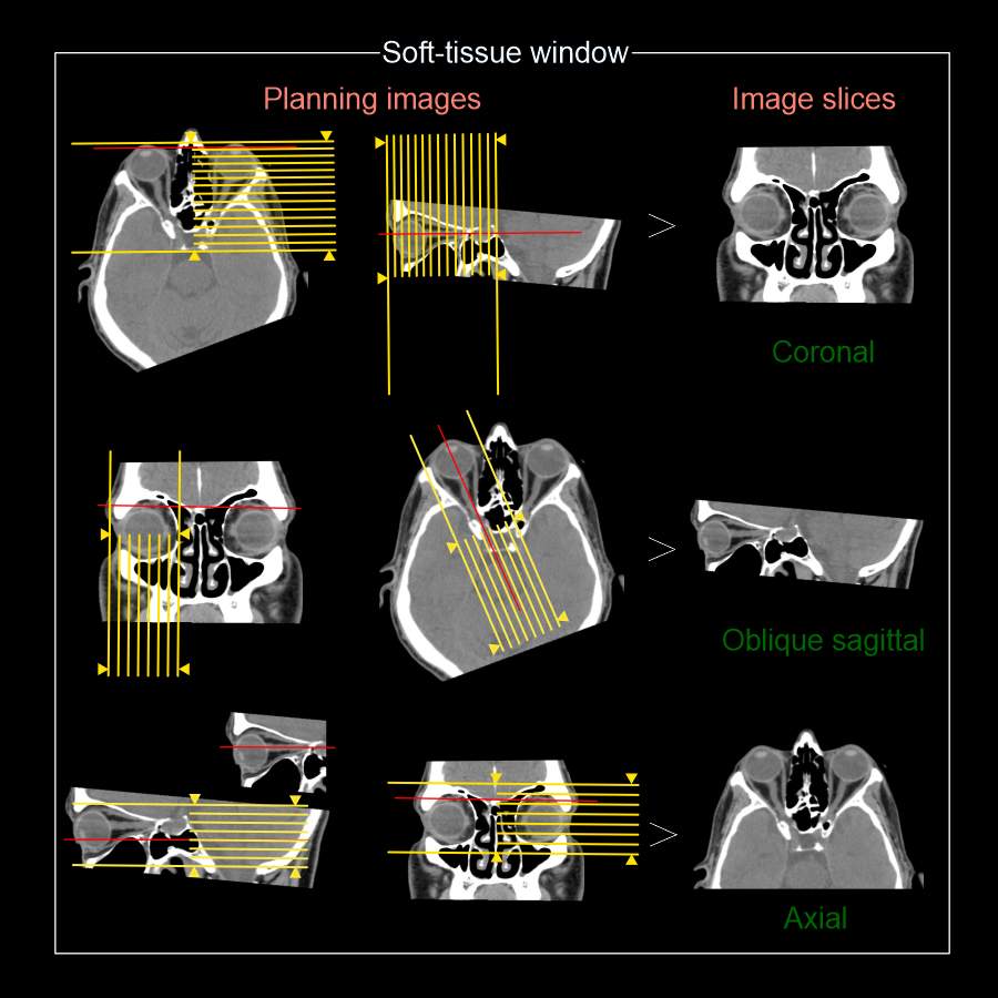
Oblique sagittal images in soft-tissue window to show the optic nerve. This is achieved by planning the oblique sagittal slices parrel to the optic nerve seen in the axial plane. Reconstruct separate images both right and left optic nerves.
- Oblique sagittal images in soft-tissue window to show both optic nerves.
Explanation: this is achieved by using axial plane to plan oblique-sagittal slices parrel to the optic nerve.
Reference
- Ashley H. Aiken, MD, Chair, Paul M. Bunch, MD, & Kavita K. Erickson, MD. (2021). ACR–ASNR–SPR Practice parameter for the performance of computed tomography (CT) of the extracranial head and neck.Retrieved from www.gravitas.acr.org.
- Radiology Service. (2023, June 30). CT- Orbits non contrast. Retrieved from www.starship.org.nz
- Tawfik, H. A., Abdelhalim, A., & Elkafrawy, M. H. (2012). Computed tomography of the orbit - A review and an update. Saudi journal of ophthalmology: official journal of the Saudi Ophthalmological Society, 26(4), 409–418. https://doi.org/10.1016/j.sjopt.2012.07.004
- Cellina, M., Cè, M., Marziali, S., Irmici, G., Gibelli, D., Oliva, G., & Carrafiello, G. (2022).Computed tomography in traumatic orbital emergencies: a pictorial essay-imaging findings, tips, and report flowchart.Insights into imaging, 13(1), 4. https://doi.org/10.1186/s13244-021-01142-y
- Romanyukha, A., Nzitunga, P. S., & Dolcet, A. (2022, April 28). CT patient positioning plays key role in radiation dose reduction. www.auntminnie.com.