CT Contrast chest scan
Last updated May 13, 2026
Similar expressions
CECT chest, Chest contrast
Introduction
CT Chest contrast scan is mainly used to diagnose conditions related to thoracic cavity, including lung nodules, mediastinal lesions and infections. This is routinely requested as further assessment for abnormalities recognized in chest x-rays.
Patient preparation
- Explain the procedure clearly and kindly.
- Remove metals related to the scanning area (underwear, necklaces).
- Practice breathing technique – breath in and hold during the scan.
- Check contraindications for contrast media administration and radiation exposure.
- Place an Intra venous (IV) cannula in a stable vein for contrast injection – pink-20G cannula.
- Explain the burning sensation during contrast injection.
Patient positioning
- Position the patient in supine and feet first on the imaging couch or table.
- Center the scanning area in the scanner iso-center [6].
Explanation: This reduces overall radiation exposure and increases image quality.
- Both hands are raised above the head, positioning aide or a pillow can be placed under the hands for comfort.
Explanation: raising hands above the head reduces streak artifacts, and also avoids unnecessary radiation exposure to the hands.
- Keep the arm with the IV cannula strait.
Explanation: to facilitate contrast flow.
- Plan the scan starting point at the lower neck.
Scan planning
Scan has two phases, which are pre-contrast and arterial. It is hard to define an accurate time for the chest arterial phase, which is nearly 25 seconds from contrast injection, because it changes according to the patient’s ejection fraction. So, triggering method can be used to initiate a timely arterial phase by assessing patient’s real-time arterial enhancement. Please reach our thoracic aortogram article for more information about bolus tracking.
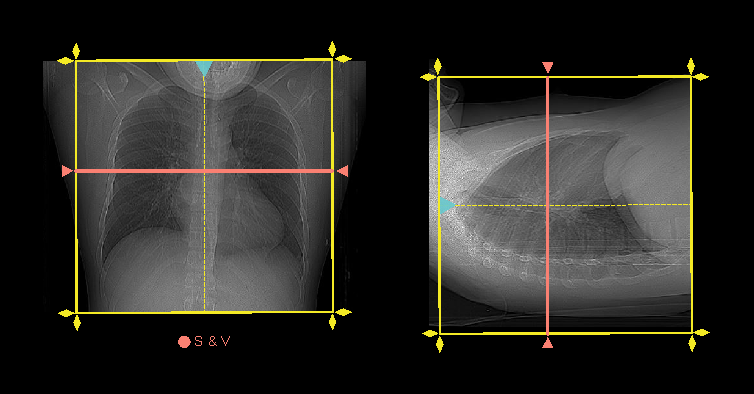
- Plan both scan slabs to cover from lung apex to posterior costophrenic sulci.
- Acquire both phases under suspended full inspiration.
- Place scan and view (S & V) slice below carina.
- Place triggering region of interest (ROI) in the descending aorta.
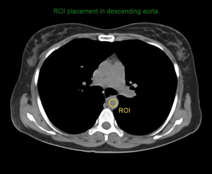
- Program the threshold value to 160 HU.
- Set S & V initiation delay to 10 seconds.
- Place zero delay time for arterial scan.
Intravenous (IV) contrast infusion
- Inject 65-70ml (65kg body weight) of iodinated contrast at a rate of 3ml/s.
Post-processing
- Axial, coronal and sagittal images with <3 mm slice thickness in lung window (WW: 1500, WL: -500).
- Axial, coronal and sagittal images with <5mm slice thickness in soft-tissue window (WW: 400,WL:40).
- Axial images with <3mm slice thickness in bone window (WW: 3500, WL: 350).
Post processing plays a key role in diagnosis, and some image categories are special to demonstrate specific abnormalities. Please reach our HRCT chest and chest non contrast articles for more information.
Reference
- Mannudeep K. S. Kalra, MD, Chair, Jessica Kurian MD, & Satinder Singh, MD, FSABI. (2023). ACR–SABI–SPR–STR Practice parameter for the performance of thoracic computed tomography (CT). Retrieved from www.gravitas.acr.org.
- Bhalla, A. S., Das, A., Naranje, P., Irodi, A., Raj, V., & Goyal, A. (2019). Imaging protocols for CT chest: A recommendation. The Indian journal of radiology & imaging, 29(3), 236–246. https://doi.org/10.4103/ijri.IJRI_34_19
- Hochhegger, B., Rottenfusser, R., & Marchiori, E. (2017). When is the use of contrast media in chest CT indicated?. Jornal brasileiro de pneumologia : publicacao oficial da Sociedade Brasileira de Pneumologia e Tisilogia, 43(5), 400. https://doi.org/10.1590/S1806-37562017000000179
- Kul, M., Kuru Öz, D., Gürsoy Coruh, A., Özalp Ateş, F., Gülpınar, B., Uzun, Ç., & Atasoy, K. Ç.(2022). Biphasic split-bolus injection protocol for routine contrast-enhanced chest CT: comparison with conventional early-phase single bolus technique. The British journal of radiology, 95(1134), 20210775. https://doi.org/10.1259/bjr.20210775
- Yang, R., Yan, Y., Wang, Y., Liu, X., & Su, X. (2017). Plain and contrast-enhanced chest computed tomography scan findings of pulmonary cryptococcosis in immunocompetent patients. Experimental and therapeutic medicine, 14(5), 4417–4424. https://doi.org/10.3892/etm.2017.5096
- Romanyukha, A., Nzitunga, P. S., & Dolcet, A. (2022, April 28). CT patient positioning plays key role in radiation dose reduction.www.auntminnie.com.