4D CT Parathyroid Protocol
Last updated June 27, 2026
By Radiohelp Staff
Similar expressions
CT Parathyroid 4D neck scan/ CT contrast 4D neck scan/ CT 4D neck
Introduction
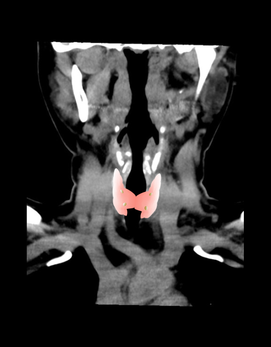
4D Parathyroid CT is mainly used to diagnose abnormalities associated with the parathyroid gland, Specially, localizing parathyroid adenomas for surgeries. As it’s in the name, scan has 4 dimensions including 2D images, time (phases) and multiple planes.
Patient preparation
- Explain the procedure kindly and clearly.
- Remove metal related to the neck area (necklaces, undergarments).
- Instruct the patient to be steady and not to swallow during the procedure.
- Ask to breath quietly during the procedure.
- Assess the ability to administer contrast media and expose radiation.
- Place a cannula in a stable vein for contrast injection – pink-20G cannula.
Patient positioning
- Position in head-first and supine.
- Extend the neck slightly.
Explanation: reduces streak artifacts or beam hardening artifacts due to the lower jaw. Same results can be achieved by angulating CT gantry.
- Position the shoulders in a pulled down position and arms next to the body.
Explanation: reduces streak artifacts or beam hardening artifacts due to wide shoulders. Placing a cushion under mid-upper thorax moves shoulders posteriorly, and helps to reduce streak artifacts at the root of the neck [2].
- Hand, which is used to infuse contrast media, is placed straight to facilitates the contrast flow.
- Center the scanning area in the scanner iso-center [5].
Explanation: this reduces overall radiation exposure and increases image quality.
- Plan the scout starting point at the nipple level.
Scan planning
- Three to four phases are performed: pre-contrast, arterial phase, venous phase and/or Delayed phase.
- For all phases, plan the scan slab to cover from the skull base to the aortopulmonary window or carina.
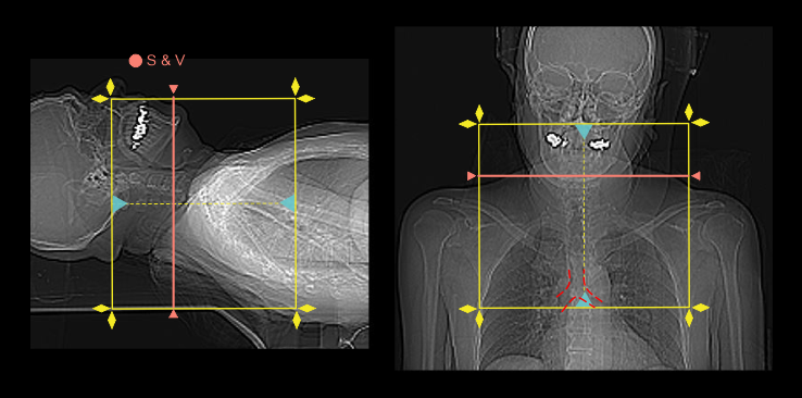
Triggering method or bolus tracking method can be used to initiate the arterial phase because arterial enhancement can be viewed in real-time from the scan and view (S & V), without being delayed to acquire a mixture of arterial and venous enhancements. Additionally, starting the arterial phase in manual is better due to the difficulty of placing a ROI in the small carotid arteries, particularly, in the non-contrast S & V.
- Place the scan and view (S & V) slice at the mid part of the neck.
- Set S & V initiation time to 5 seconds from the contrast injection.
- Plan the scan direction for arterial phase from head to foot.
Explanation: this direction avoids delayed acquisition in superior region of the neck visualizing veins, and evades streak artifacts at the root of the neck when there is undiluted contrast media in brachiocephalic and subclavian veins.
- Plan the scan direction for venous phase from foot to head with a 2 second delay.
Explanation: this is to avoid over delay of venous phase because scan is started at the point where the arterial phase ends, skipping the time it takes to move to skull base.
- Set a 3-5 min delay time for the delayed phase.
- Start arterial phase manually without any delay when there is adequate carotid enhancement – please review our neck contrast article for images.
Intravenous (IV) contrast administration
- Pressure injector is used to inject 50-60 ml of iodinated contrast media at a rate of 3ml/s (body weight of 60kg).
- 40-50ml saline chase or flush is used.
Post-processing
- Axial, coronal and sagittal images in soft-tissue window (WW: 400, WL:40) without exceeding 3mm slice thickness - refer neck contrast article for images.
- Additionally, a suitable image in bone (WW: 3500, WL:350) window, displayed in any plane, without exceeding 3mm slice thickness.
Reference
- Ashley H. Aiken, MD, Chair, Paul M. Bunch, MD, & Kavita K. Erickson, MD. (2021). ACR–ASNR–SPR Practice parameter for the performance of computed tomography (CT) of the extracranial head and neck.Retrieved from www.gravitas.acr.org.
- Harvey, G. D., Mayer, D. P., & Radecki, P. D. (1984). Simplified patient positioning to reduce beam hardening in CT of the lower neck. AJNR. American journal of neuroradiology, 5(6), 796.
- Nagano, S. Y. M., Bitencourt, A. G. V., Torres, I. D. C. G., & Porto, G. C. L. M. (2021). Four-dimensional computed tomography protocol for preoperative evaluation of the parathyroid glands and its correlations with other imaging methods: a pictorial essay. Radiologia brasileira, 54(3), 193–197. https://doi.org/10.1590/0100-3984.2020.0056
- Bunch PM, Randolph GW, Brooks JA, George V, Cannon J, Kelly HR. Parathyroid 4D CT: What the Surgeon Wants to Know.Radiographics. 2020 Sep-Oct;40(5):1383-1394. doi: 10.1148/rg.2020190190. Epub 2020 Jul 17. PMID: 32678698.
- Romanyukha, A., Nzitunga, P. S., & Dolcet, A. (2022, April 28). CT patient positioning plays key role in radiation dose reduction.www.auntminnie.com.
- Kelly, H. R., Hamberg, L. M., & Hunter, G. J. (2014). 4D-CT for preoperative localization of abnormal parathyroid glands in patients with hyperparathyroidism: accuracy and ability to stratify patients by unilateral versus bilateral disease in surgery-naive and re-exploration patients. AJNR. American journal of neuroradiology, 35(1), 176–181. https://doi.org/10.3174/ajnr.A3615