CT Cervical Spine
Last updated June 27, 2026
By Radiohelp Staff
Similar expressions
CT cervical spine/ CT C spine
Introduction
Cervical CT scan is mainly used to assess cervical spine injures such as fractures. It also helps to diagnose spine neoplasms, congenital abnormalities and implant checks.
Patient preparation
- Explain the examination clearly and kindly.
- Ask to remove radio opaque items such as ear rings, necklaces, underclothing and piercing related to cervical region.
Patient positioning
- Position the patient in head-first, supine and in the iso-center.
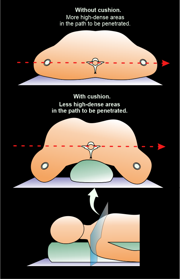
- Keep shoulders in a pulled down position and arms next to the body.
Explanation: this reduces streak artifacts or beam hardening artifacts due to wide shoulders. Placing a cushion under mid-upper thorax moves shoulders posteriorly, and helps to reduce streak artifacts at the root of the neck.
- Plan the scan start point at the level of clavicles and set scan direction outward the gantry.
Scan planning
- Plan the scan slab to cover from skull base (or C1) to first thoracic vertebra (T1).
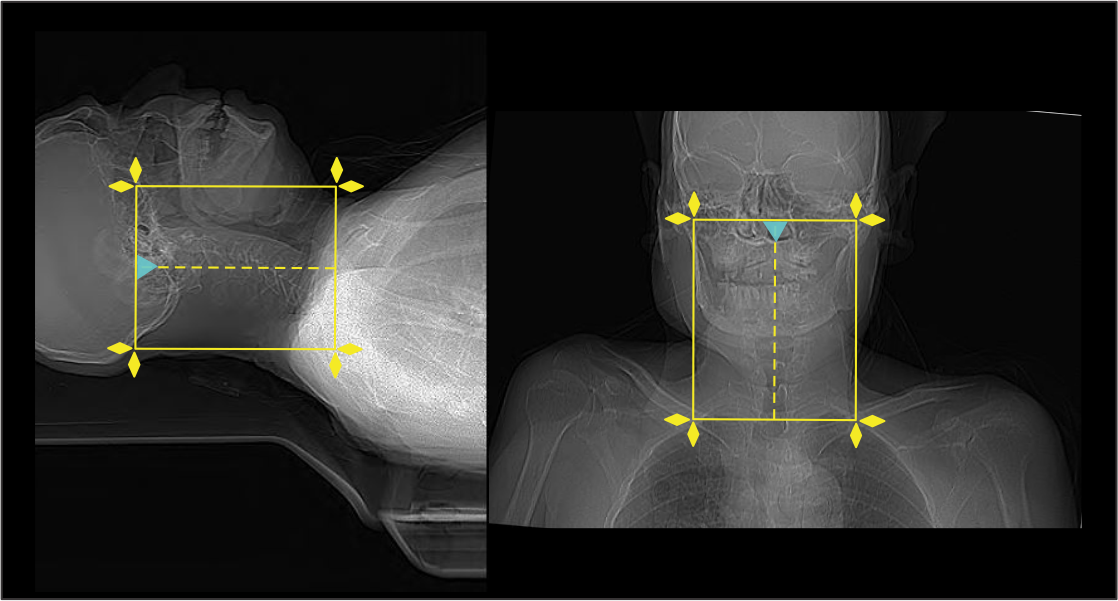
- Reduce the field of view (FOV) as small as appropriate.
Explanation: smaller FOV increases geometric resolution of the image.
Post-processing
- Sagittal and coronal images with ≤ 2 mm slice thickness in bone window (WW: 3500, WL: 350).
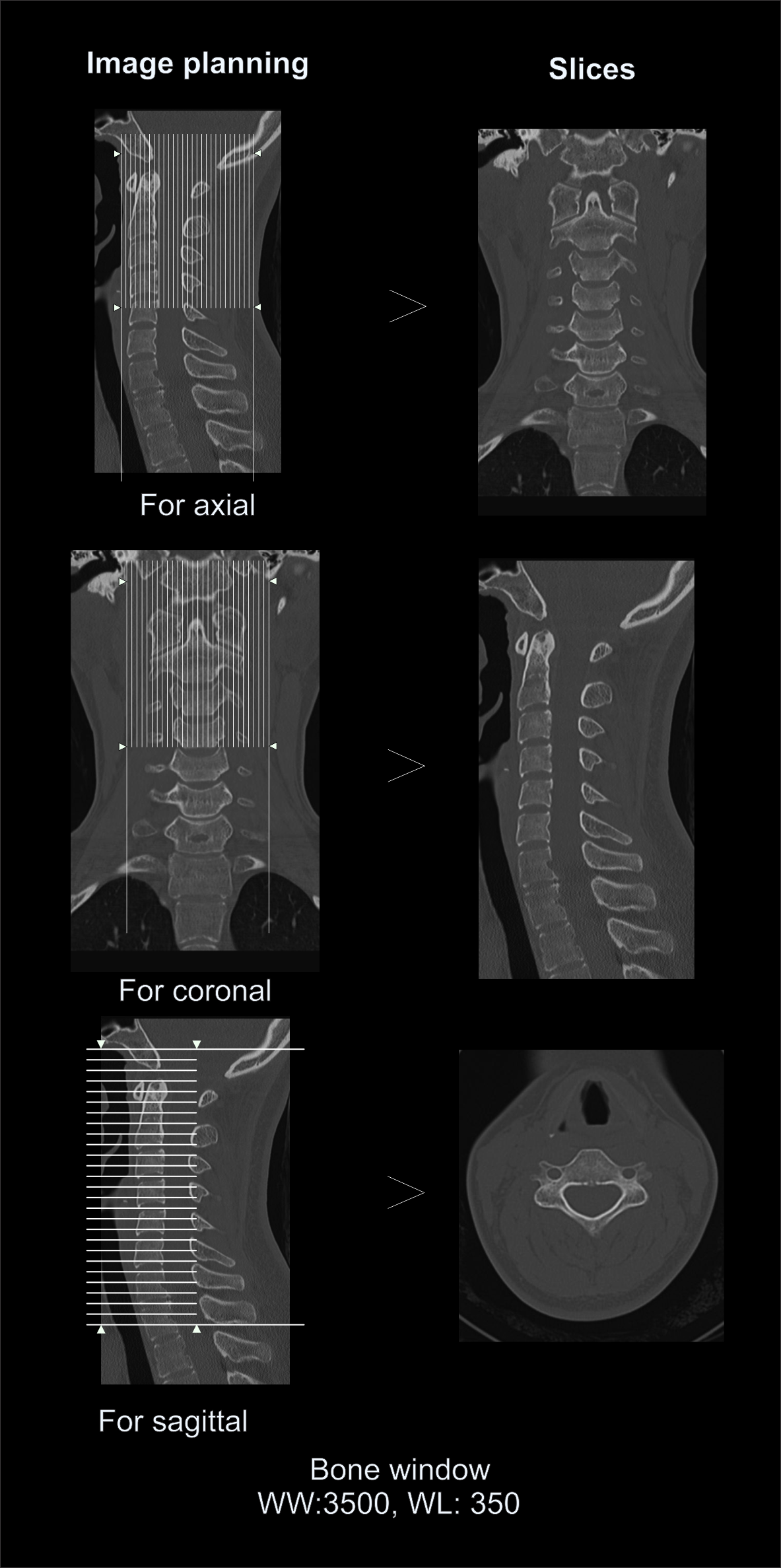
- Contagious axial slices in bone window and soft-tissue window (WW:500, WL:50) with ≤ 2mm and ≤ 3mm slice thicknesses respectively.
- Oblique reformats perpendicular to the long axis of the neural foramina on both sides.
Explanation: for the evaluation of neural foraminal stenosis.
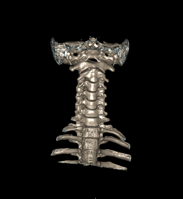
- Additionally, 3d images to show Cervical-spine.
Reference
- Harvey GD, Mayer DP, Radecki PD. Simplified patient positioning to reduce beam hardening in CT of the lower neck.AJNR Am J Neuroradiol. 1984 Nov-Dec;5(6):796. PMID: 6437182; PMCID: PMC8333654.
- Lubdha M. Shah, MD, Chair, Kristine A. Blackham, MD, & Kavita K. Erickson, MD. (2022). ACR–ASNR–ASSR–SPR practice parameter for the performance of computed tomography (CT) of the spine. Retrieved from www.gravitas.acr.org.
- Tozakidou M, Yang SR, Kovacs BK, Szucs-Farkas Z, Studler U, Schindera S, Hirschmann A. Dose-optimized computed tomography of the cervical spine in patients with shoulder pull-down: Is image quality comparable with a standard dose protocol in an emergency setting? Eur J Radiol. 2019 Nov;120:108655. doi: 10.1016/j.ejrad.2019.108655. Epub 2019 Sep 12. PMID: 31542699.
- Derakhshan, A., Lubelski, D., Steinmetz, M. P., Benzel, E. C., & Mroz, T. E. (2015). Utility of Computed Tomography following Anterior Cervical Diskectomy and Fusion. Global spine journal, 5(5), 411–416. https://doi.org/10.1055/s-0035-1554773
- Riahi, H., Mechri, M., Barsaoui, M., Bouaziz, M., Vanhoenacker, F., & Ladeb, M. (2018). Imaging of Benign Tumors of the Osseous Spine.Journal of the Belgian Society of Radiology, 102(1), 13.https://doi.org/10.5334/jbsr.1380
- Romanyukha, A., Nzitunga, P. S., & Dolcet, A. (2022, April 28). CT patient positioning plays key role in radiation dose reduction.www.auntminnie.com.
- Okamoto, A., Takeshima, Y., Yokoyama, S., Nishimura, F., Nakagawa, I., Park, Y. S., & Nakase, H. (2022). Prevalence and Clinical Impact of Cervical Facet Joint Degeneration on Degenerative Cervical Myelopathy: A Novel Computed Tomography Classification Study. Neurospine, 19(2), 393–401. https://doi.org/10.14245/ns.2143258.629