CT Angiography lower limb arteries
Last updated June 03, 2026
By Radiohelp Staff
Similar expressions
CECT lower limb angio/ CECT peripheral angiogram lower limb
Introduction
CT lower limb angiogram is a contrast enhanced study that helps to diagnose complications associated with the peripheral vascular system of lower limbs, including vessel stenosis, congenital abnormalities and arterial occlusions.
Patient preparation
- Assess suitability to expose radiation and administer iodinated contrast media intravenously.
- Remove metallic objects in the interested region.
- Practice 'breath in and hold' technique.
- Place an 18-gauge cannula in a stable vein.
Patient positioning
- Position in feet-first supine.
- Place both legs flat on the table and immobilize them.
- Keep the interested region in the iso-center of the scanner.
Explanation: this reduces radiation dose to patient and improves image quality.
- Raise hands and keep them beside head.
Explanation: avoids streak artifacts to chest and abdominal area. It also reduces radiation exposure to patient.
Important: this scan is one of the lengthiest body-coverage CT scans, so move the couch to check the moving range, and avoid tube tangling or accidental stop due to limited couch range.
Scan planning
There are two ways to perform this study. In one method, only the arterial phase is acquired, and in the other, both pre and post contrast phases are achieved.
Method 1 – only arterial phase
Radiation dose is lower compared to the other, but there is more post processing which is time consuming.
- Plan the scan slab to cover from diaphragm to ends of lower limbs (toes).
- Triggering or bolus tracking method is used – refer thoracic aortogram article for more details.
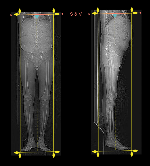
- Keep scan and view (S & V) slice just above diaphragm.
- Plan 10 seconds delay from contrast injection to S & V.
Explanation: contrast reach nearly after 20 seconds in to abdominal aorta.
- Place 10 seconds delay to start arterial phase.
Explanation: because time is needed for the contrast to reach distal areas.
Method 2 – pre and arterial phases
Postprocessing is less in this method because pre contrast phase is subtracted from arterial phase to get the only arterial image. Two phases may be compared to identify vascular calcifications. Radiation dose is higher in this method due to dual phase acquisition. Scan coverage and planning is same as method 1. It is important to keep ideal dimensions for both pre and arterial phases, otherwise subtraction is not possible.
Intra venous (IV) contrast administration
- Inject 95 ml (body weight 80kg) of iodinated contrast media at a rate of 4.5 ml/s with the help of a pressure injector.
- Inject 70 ml of saline flush just after completing the contrast volume.
Explanation: flushes remaining contrast in veins of the injected hand and helps to maintain contrast flow for a longer time.
Important: when administering IV contrast, factors such as injection rate, contrast volume, and infusion technique might be changed according to the patient body weight, patient condition, scan coverage, departmental protocol and CT scanner model. So, an experienced user is essential here for the safety of the patient.
Post-processing
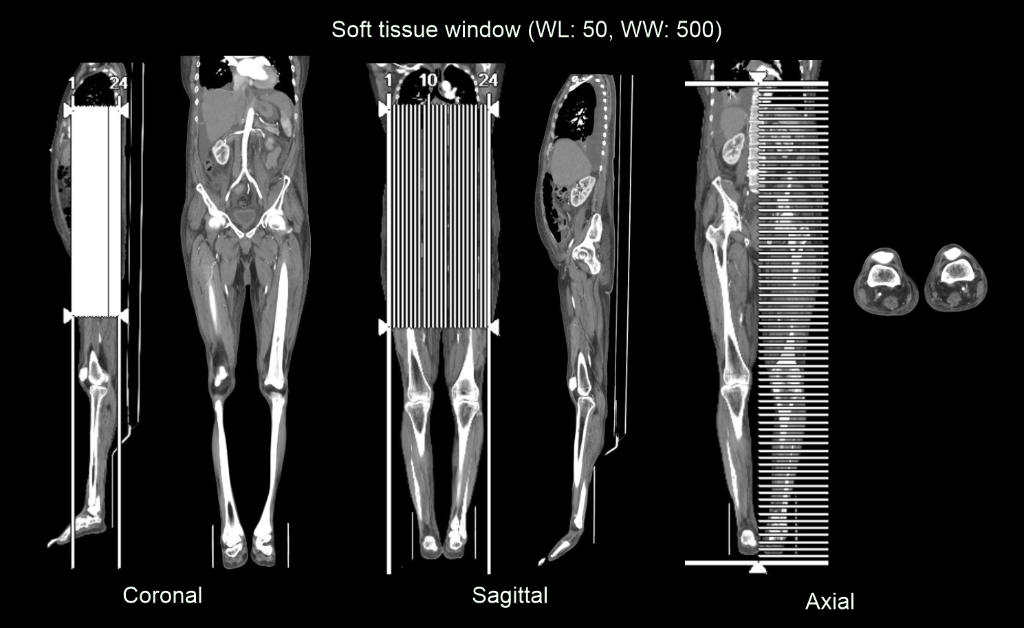
- Axial, sagittal and coronal images in both bone (WW: 3500, WL: 350) and soft tissue window (WW: 500, WL: 50) with ≤ 2mm slice thickness.
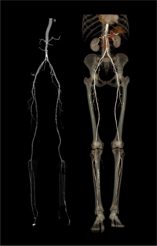
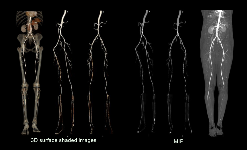
- 3D surface shaded images and MIP (maximum intensity projection) images to clearly demonstrate arteries of lower limbs.
Reference
- Rotzinger DC, Lu TL, Kawkabani A, Marques-Vidal PM, Fetz G, Qanadli SD. Computed Tomography Angiography in Peripheral Arterial Disease: Comparison of Three Image Acquisition Techniques to Optimize Vascular Enhancement-Randomized Controlled Trial.Front Cardiovasc Med. 2020 Apr 28;7:68. doi: 10.3389/fcvm.2020.00068. PMID: 32411728; PMCID: PMC7198850.
- Steven S. Raman, MD, Chair, Dorothy Gilbertson, MD, & Charles White, MD. (2021). ACR–NASCI–SIR–SPR Practice parameter for the performance and interpretation of body computed tomography angiography (CTA).Retrieved from www.gravitas.acr.org.
- Horehledova, B., Mihl, C., Milanese, G., Brans, R., Eijsvoogel, N. G., Hendriks, B. M. F., Wildberger, J. E., & Das, M. (2018). CT Angiography in the Lower Extremity Peripheral Artery Disease Feasibility of an Ultra-Low Volume Contrast Media Protocol. Cardiovascular and interventional radiology, 41(11), 1751–1764. https://doi.org/10.1007/s00270-018-1979-z
- Keddie D, Abdulrehman Y, Shiau G. Reporting lower extremity CT angiography for treatment planning.Diagn Interv Imaging. 2022 Sep;103(9):387-393. doi: 10.1016/j.diii.2022.06.010. Epub 2022 Jul 14. PMID: 35843841.
- Gakhal MS, Sartip KA. CT angiography signs of lower extremity vascular trauma. AJR Am J Roentgenol. 2009 Jul;193(1):W49-57. doi: 10.2214/AJR.08.2011. PMID: 19542383.
- Shetty AS, Hoegger MJ, Rajput MZ, Itani M, Nhan DT, Perez AA, Webb IG, Wiltshire JP, Feister KF, Ballard DH, Raptis D, Raptis CA, Mellnick VM, Lanier MH, Naeem M, Duwayri Y, Tsai R. Aortoiliofemoral Lower Extremity CT Angiography. Radiographics.2025 Oct;45(10):e240272. doi: 10.1148/rg.240272. PMID: 40906585.