CT Whole Spine Scan
Last updated June 19, 2026
By Radiohelp Staff
Similar expressions
CT Whole spine/ CT full spine
Introduction
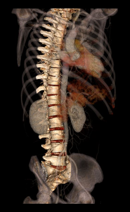
CT Whole spine protocol is used for the assessment of spine pathologies including scoliosis, Kyphosis, pathological fractures and compression fractures (more spine related pathologies). Moreover, scan images are useful to measure the pedicle width for pedicle screw placement surgeries, and pre and post surgical evaluation.
Patient preparation
- Explain the examination clearly and kindly.
- Ask to remove radio opaque items such as hair clips, necklaces, earrings and underclothing from head to end of pelvic bone.
Patient positioning
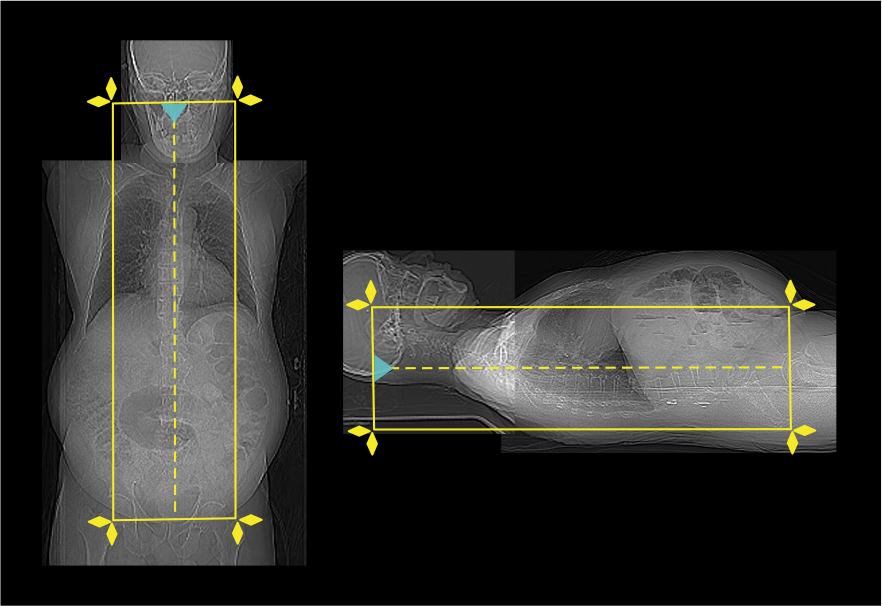
- Position in head-first, supine and in the iso-center.
Explanation: patient in the iso-center reduces radiation exposure and improves image quality.
- Both hands are raised above the head.
Explanation: this reduces streak artifacts due to hands.
- Plan the scout start point at nasion and set scan direction inward the gantry.
Scan planning
- Plan the scan slab to cover from C1 to end of sacrum.
- Reduce the field of view (FOV) as small as appropriate.
Explanation: smaller FOV increases the geometric resolution of the image.
Post-processing
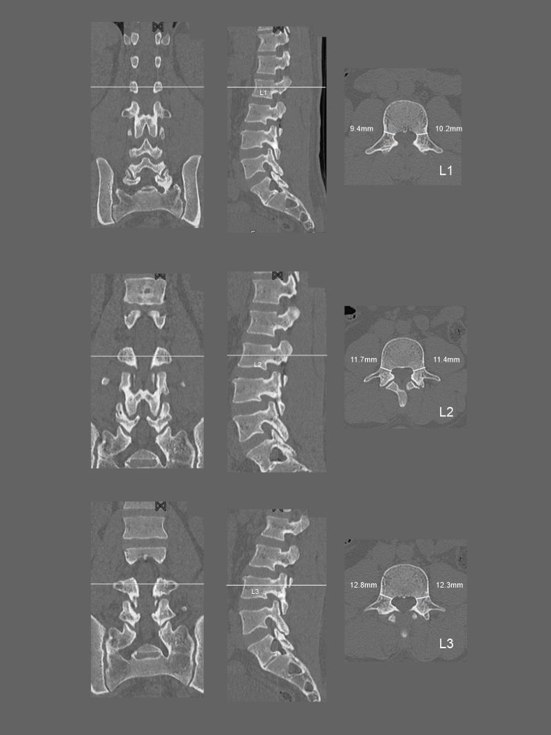
- Sagittal and coronal images with ≤ 2 mm slice thickness in bone window.
- Curved multi-planar reconstruction (curved MPR) is useful when there are abnormal curvatures in the spine.
- Contagious axial slices in bone window (WW: 3500, WL: 350) and soft-tissue window (WW:500,WL:50) with ≤ 2mm and ≤ 3mm slice thicknesses respectively.
- Pedicle width of vertebrae from C1 to L5 if the purpose of the scan is pre surgical planning for scoliosis correction.
- Additionally, 3d images to show the full spine.
Reference
- Lubdha M. Shah, MD, Chair, Kristine A. Blackham, MD, & Kavita K. Erickson, MD. (2022). ACR–ASNR–ASSR–SPR practice parameter for the performance of computed tomography (CT) of the spine. Retrieved from www.gravitas.acr.org.
- Ahn, Tae-Keun & Bourret, Stephane & Thompson, Wendy & Roscop, Cecile & Cloché, Thibault & Lehuec, Jean-Charles. (2020). Full Length Spine CT and MRI. International Journal of Clinical Medicine. 11. 270-281. 10.4236/ijcm.2020.115028.
- Sakti YM, Lanodiyu ZA, Ichsantyaridha M, Wijanarko S, Filza MR, Taufan T, Susanto DB, Tampubolon YO, Baskara AANN, Nurshal AA, Mustofa FD, Rosfadilla A, Magetsari R, Rukmoyo T. Pedicle morphometry analysis of main thoracic apex adolescent idiopathic scoliosis. BMC Surg. 2023 Feb 9;23(1):34. doi: 10.1186/s12893-022-01877-5. PMID: 36759804; PMCID: PMC9912543.
- Shi H, Zhu L, Ma J, Zhu YC, Wu XT. The accuracy of a novel pedicle screw insertion technique assisted by a special angular scale in the subaxial cervical spine using lateral mass as a reference marker.J Orthop Surg Res. 2020 Nov 23;15(1):551. doi: 10.1186/s13018-020-02054-1. PMID: 33228707; PMCID: PMC7681976.
- Menger, R. P., Storey, C. M., Nixon, M. K., Haydel, J., Nanda, A., & Sin, A. (2015). Placement of C1 Pedicle Screws Using Minimal Exposure: Radiographic, Clinical, and Literature Validation. International journal of spine surgery, 9, 43. https://doi.org/10.14444/2043
- Romanyukha, A., Nzitunga, P. S., & Dolcet, A. (2022, April 28). CT patient positioning plays key role in radiation dose reduction.www.auntminnie.com.