CT Cisternogram
Last updated June 27, 2026
By Radiohelp Staff
Similar expressions
CT Cisternography/ CECT Cisternogram
Introduction
CT Cisternography is a contrast study used to diagnose abnormal cerebrospinal fluid (CSF) leaks-mainly into areas such as paranasal sinuses, nasal cavity (CSF rhinorrhea), middle ear cavity (CSF otorrhea), subarachnoid space and mastoid air cells.
Patient preparation
- Remove hair clips, ear rings or any removable metal in the exposing area.
- Explain the procedure clearly.
- Prepare the patient for contrast administration and lumbar puncture.
Contrast administration:
- Fluoroscopy guided lumbar puncture is performed by a Radiologist.
- Iodinated contrast media is administered into the thecal sac.
- Table is tilted to make the patient’s head down.
Explanation: this position helps to move contrast media towards the head.
- Patient is transferred to the CT room after a successful lumbar puncture and contrast administration.
Patient positioning
Patient positioning may change according to the condition of the patient.
- Head-first-prone position is suitable if the patient is leaking CSF from the nasal cavity (CSF rhinorrhea).
Explanation: position the patient to promote active leakage of CSF (slight elevation of the pelvic area), sneezing can also be an action to promote CSF leakage.
- Center the scanning area in the scanner iso-center [2].
Explanation: this reduces overall radiation exposure and increases image quality.
- Radio-lucent pad can be kept under the forehead to make room between the table surface and face.
Scan planning
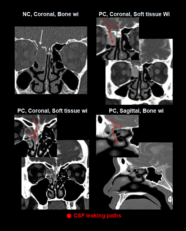
Non-contrast CT head may help to identify contrast leaking areas in comparison with thepost-contrast images.

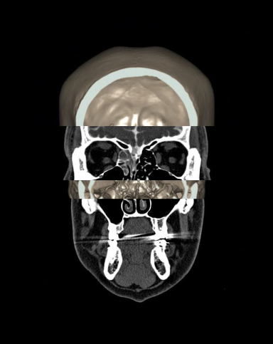
- Plan the scan starting point at the angle of the mandible (patient in prone and head first position).
- Plan the scan slab to cover from the skull vertex to mandible.
Post-processing
- Axial, sagittal and coronal images in bone (WW:4500, WL: 450) and soft-tissue (WW:400, WL: 40) windows, with slice thickness lower than 2mm.
Explanation: these images should clearly demonstrate CSF leaks.
Please refer to our Brain contrast and Sinus article for MPR planning and images.
Reference
- Amrhein, T. J., Goldman-Yassen, Adam MD, & Ali, Saad MD. (2024). ACR–ASNR–SPR Practiceparameter for the performance of myelography and cisternography. Retrieved from gravitas.acr.org
- Romanyukha, A., Nzitunga, P. S., & Dolcet, A. (2022, April 28). CT patient positioning plays key role in radiation dose reduction. www.auntminnie.com.
- Hablas, L.T., Ammar, A.M. & Elnagar, R.M. CSF rhinorrhea: non-contrast CT, contrast-enhanced CT cisternography or combined?. Egypt J Radiol Nucl Med 53, 201 (2022).
- Shetty PG, Shroff MM, Sahani DV, Kirtane MV. Evaluation of high-resolution CT and MR cisternography in the diagnosis of cerebrospinal fluid fistula. AJNR Am J Neuroradiol. 1998 Apr;19(4):633-9. PMID: 9576647; PMCID: PMC8337403.
- Rahalkar, Mukund & Rahalkar, Anand & Joshi, Vardhan & Sant, Kailash & Zade, Kourabhi. (2017). Computed Tomography Cisternography for the Management of Cerebrospinal Fluid Fistulae. An International Journal of Otorhinolaryngology Clinics. 9. 93-95. 10.5005/jp-journals-10003-1274.