CT scan elbow joint
Last updated April 14, 2026
Similar expressions
CT elbow/ CT EJ/ NCCT elbow
Introduction
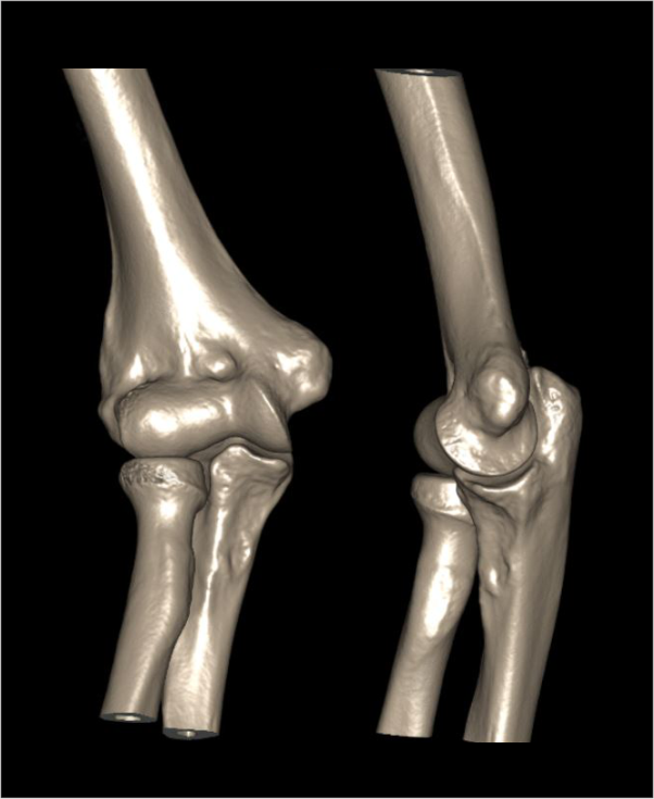
CT scan elbow joint is used to evaluate conditions ,including fractures, implant complications and neoplasm. Its ability to view structures in three dimension (3D) is remarkable for surgical planning.
Patient preparation
- Explain the procedure kindly and clearly to the patient.
- Remove radio opaque items related to the elbow area.
Patient positioning
There are several positioning methods.
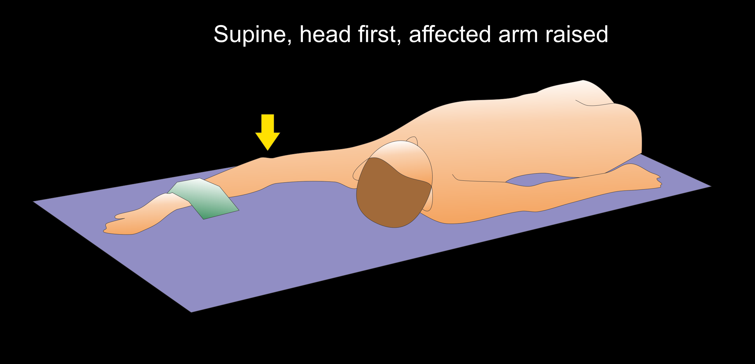
Method 1 - patient in supine, head first.
- Raise the affected arm above the head and keep the unaffected arm besides the body.
- Center the affected elbow in the iso-center.
- Extend the affected elbow-with palm facing upwards.
- Bend patient's head towards unaffected arm.
- Place a sand bag on the middle of the forearm to immobilize the affected elbow.
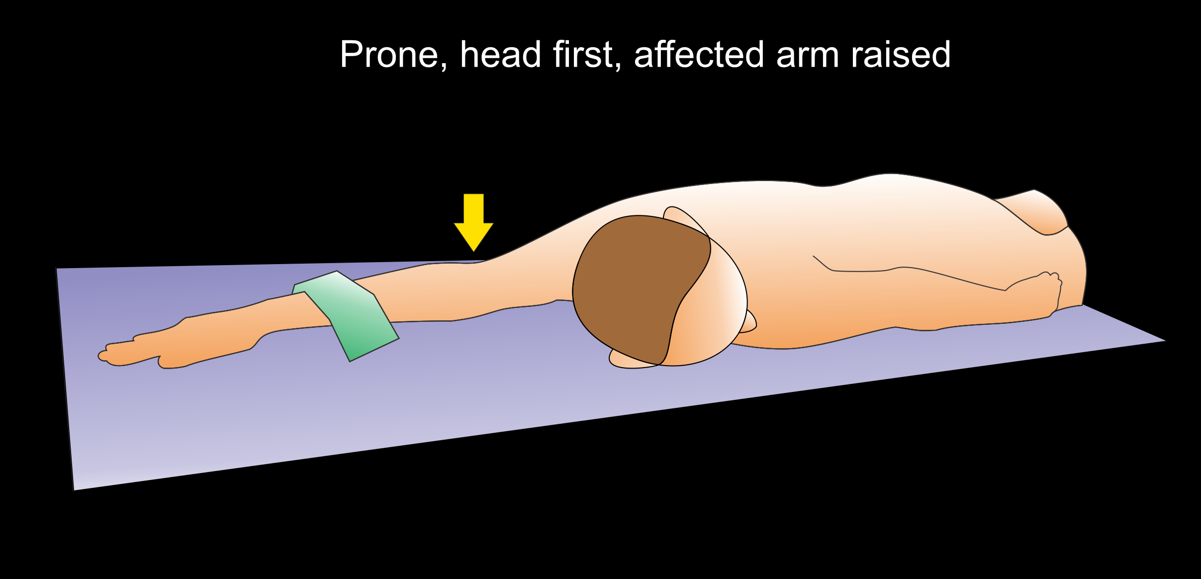
Method 2 - patient in prone, head first.
- Raise the affected arm above the head and keep the unaffected arm besides the body.
- Center the affected elbow joint in the iso-center.
- Extend the elbow-with palm facing downwards.
- Bend patient’s head towards unaffected arm.
- Place a sand bag on the middle of the forearm to immobilize the affected elbow.
Method 3 - patient in lateral decubitus.
- Position the patient in lateral decubitus on the affected side.
- Raise the affected arm above the head.
- Fully flex the neck and move the head anteriorly.
- Supinate the hand.
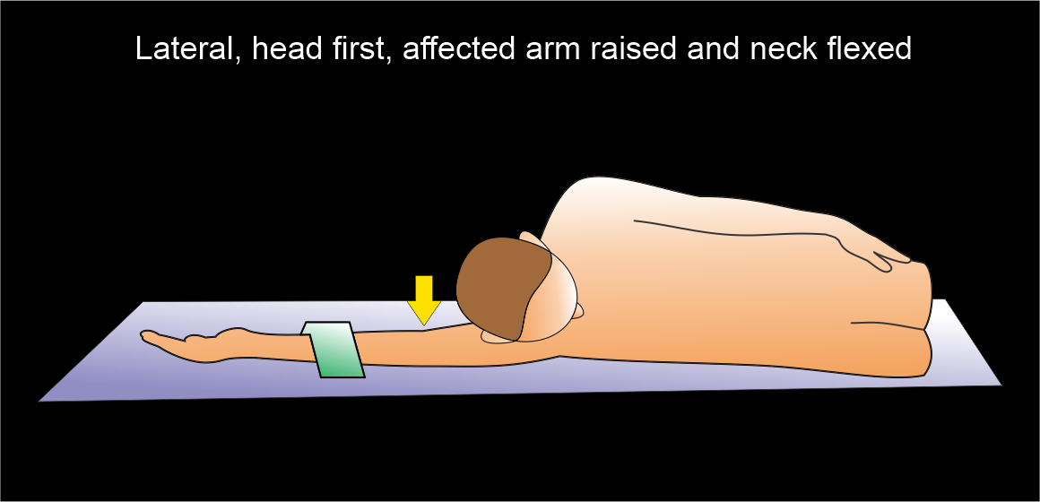
Explanation: hand in the iso-center increases image quality and lowers radiation exposure.
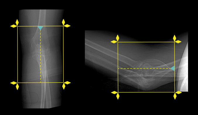
Scan planning
- Plan the scan slab to cover from the distal humeral metaphisis to proximal 1/3 of radius and ulna.
- Reduce the field of view (FOV) as appropriate to include elbow joint.
Explanation: small FOV increases geometric resolution.
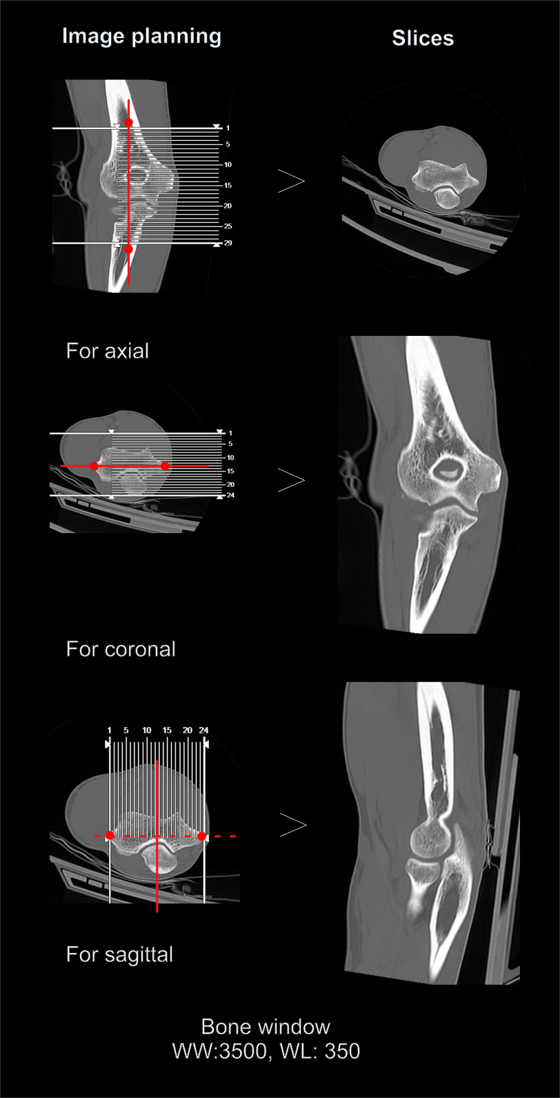
Post-processing
- Coronal, sagittal and axial images in both soft tissue (WW:500, WL:50) and bone (WW: 3500, WL: 350) window with ≤2mm slice thickness.
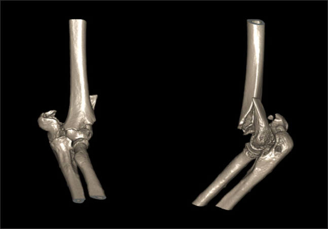
- 3d images to show pathologies clearly.
Reference
- Folco, G., Messina, C., Gitto, S., Fusco, S., Serpi, F., Zagarella, A., Gallazzi, M. B., Arrigoni, P., Aliprandi, A., Porta, M., Vitali, P., Sconfienza, L. M., & Albano, D. (2024). CT Arthrography of the Elbow: What Radiologists Should Know. Tomography (Ann Arbor, Mich.), 10(3), 415–427. https://doi.org/10.3390/tomography10030032
- Sonnow L, Salimova N, Behrendt L, Wacker FK, Örgel M, Plagge J, Weidemann F. Photon-counting CT of elbow joint fractures: image quality in a simulated post-trauma setting with off-center positioning.Eur Radiol Exp. 2023 Mar 27;7(1):15. doi: 10.1186/s41747-023-00329-w. PMID: 36967394; PMCID: PMC10040392.
- Acar K, Aksay E, Oray D, Imamoğlu T, Gunay E. Utility of Computed Tomography in Elbow Trauma Patients with Normal X-Ray Study and Positive Elbow Extension Test.J Emerg Med. 2016 Mar;50(3):444-8. doi: 10.1016/j.jemermed.2015.03.009. Epub 2015 Dec 17. PMID: 26712662.
- Liman MNP, Avva U, Ashurst JV, et al. Elbow Trauma. [Updated 2024 Apr 19]. In: StatPearls [Internet]. Treasure Island (FL): StatPearls Publishing; 2025 Jan-. Available from: https://www.ncbi.nlm.nih.gov/books/NBK542228/
- Seah, R. B., Mak, W. K., Bryant, K., Korlaet, M., Dwyer, A., & Bain, G. I. (2021). Four-dimensional computed tomography scan for dynamic elbow disorders: recommendations for clinical utility.JSES international, 6(1), 182–186. https://doi.org/10.1016/j.jseint.2021.09.013
- Romanyukha, A., Nzitunga, P. S., & Dolcet, A. (2022, April 28). CT patient positioning plays key role in radiation dose reduction.www.auntminnie.com.
- Xiao, M., Zhang, M., Lei, M., Lin, F., Chen, Y., Chen, J., Liu, J., & Ye, J. (2023). Diagnostic accuracy of ultra-low-dose CT compared to standard-dose CT for identification of non-displaced fractures of the shoulder, knee, ankle, and wrist. Insights into imaging, 14(1), 40. https://doi.org/10.1186/s13244-023-01389-7