CT Foot Scan
Last updated June 03, 2026
By Radiohelp Staff
Similar expressions
CT foot/ NCCT foot/ CT feet (both foot)
Introduction
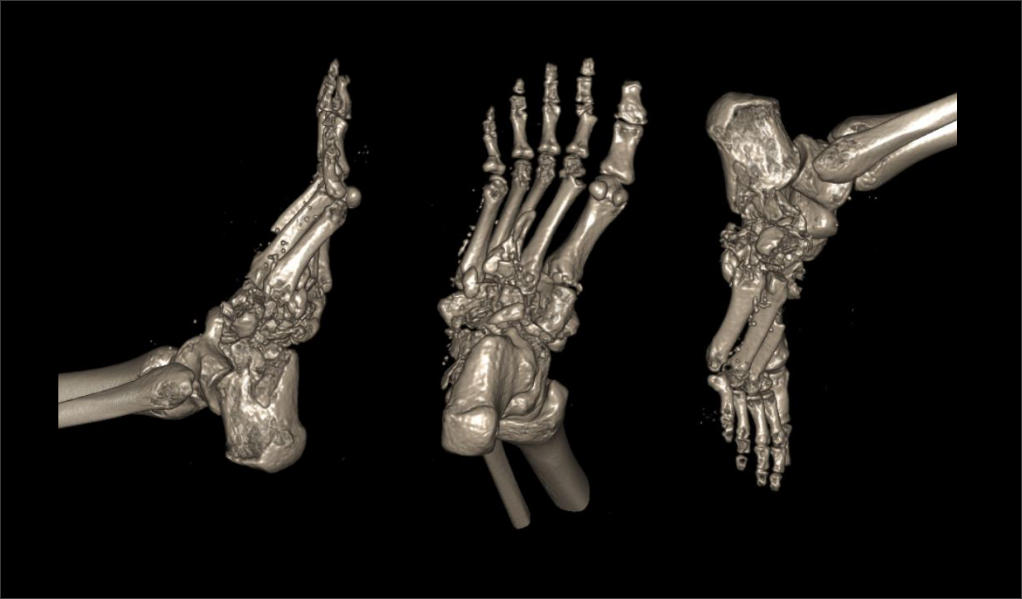
CT foot protocol is used to assess the bony structure of the foot, including fractures, neoplasms, congenital abnormalities and implants. This can provide 3D reconstruction images that helps in surgery planning.
Patient preparation
- Assess patient’s suitability to expose radiation.
- Remove removable metallic objects in the interested region.
- Ask not to move the foot or toes during the scan.
Patient positioning
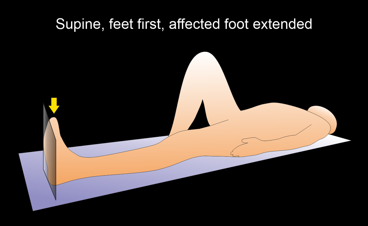
- Position in feet-first supine.
- Extend the affected leg and bent the unaffected leg at knee joint.
- Place the affected foot in the iso-center of the scanner.
Explanation: this reduces radiation dose to the patient and improves image quality.
- Position the planter aspect of the foot perpendicular to the table, a form pad can be used to support the leg.
- Place a sand bag on the mid part of the tibia to immobilize the affected limb.
Scan planning
- Plan the scan slab to cover from distal tibiofibular joint to planter aspect of the foot.
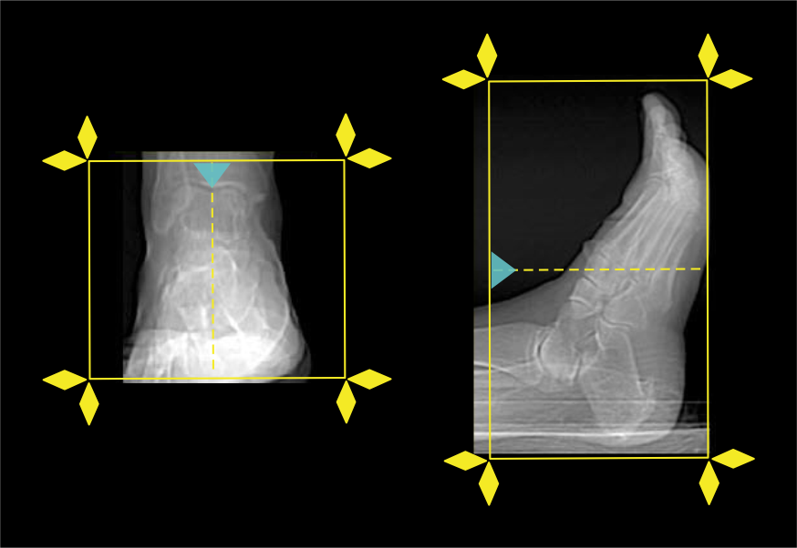
- Reduce the field of view (FOV) adequately to include the foot.
Explanation: this increases geometric resolution of the images.
Post-processing
- Axial, sagittal and coronal images in both bone (WW: 3500, WL: 350) and soft tissue window (WW:500, WL:50) with ≤ 2mm slice thickness.
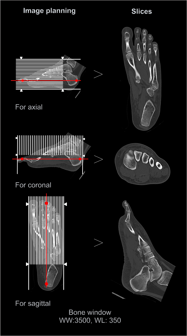
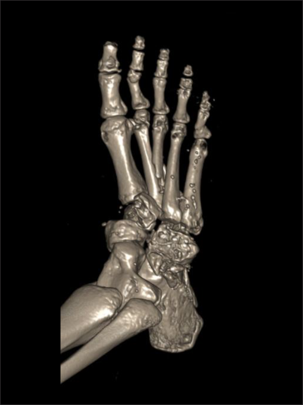
- 3D images to demonstrate pathologies clearly.
Reference
- Kalantar, S. H., Bagheri, N., Milan, N., Mehni, S. M., Oskouie, I. M., Alinia, T., & Rahimdoost, N. (2024). Evaluation of treatment planning discrepancies: CT versus plain radiographic findings in patients with foot and ankle trauma. BMC research notes, 17(1), 238. https://doi.org/10.1186/s13104-024-06902-9
- Linklater JM. Imaging of sports injuries in the foot.AJR Am J Roentgenol. 2012 Sep;199(3):500-8. doi: 10.2214/AJR.12.8547. Erratum in: AJR Am J Roentgenol. 2012 Oct;199(4):944. PMID: 22915389.
- Yoo I. R. (2020). Bone SPECT/CT of the Foot and Ankle: Potential Clinical Application for Chronic Foot Pain. Nuclear medicine and molecular imaging, 54(1), 1–8. https://doi.org/10.1007/s13139-019-00627-5