CT Knee Joint
Last updated July 01, 2026
By Radiohelp Staff
Similar expressions
NCCT knee joint/ CT KJ
Introduction
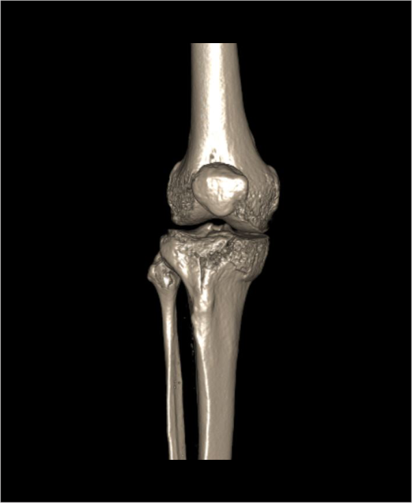
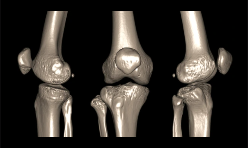
CT Knee joint is used to assess the bony structure of the knee joint, including fractures, neoplasms, congenital abnormalities and implants. This can provide 3D reconstruction images that helps in surgical planning.
Patient preparation
- Assess patient’s suitability to expose radiation.
- Remove the removable metallic objects in the interested region.
- Ask not to move the knee during the scan.
Patient positioning
- Position in feet-first supine.
- Place both legs flat on the table.
- Position the affected knee closer to the iso-center of the scanner.
Explanation: this reduces radiation dose to the patient and improves image quality.
- Position the affected knee as for a x-ray true AP projection if possible.
- Place a sand bag on the mid part of the lower leg to immobilize.
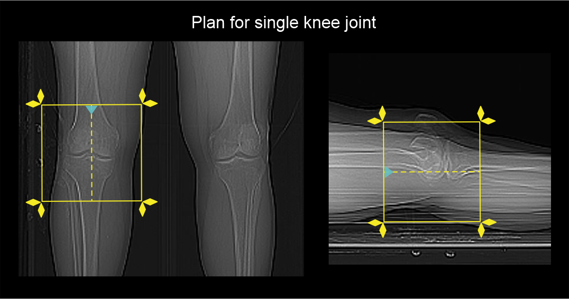
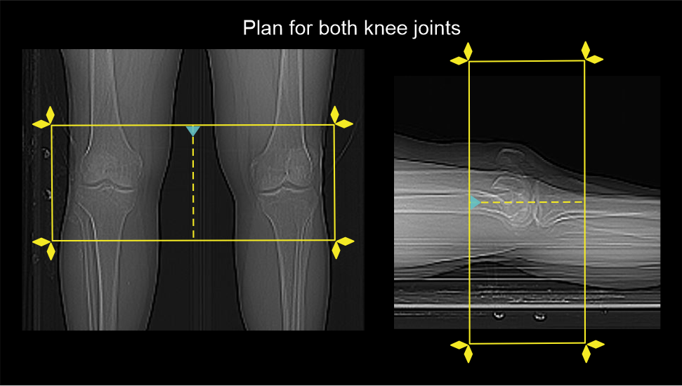
Scan planning
- Plan the scan slab to include whole patella and fibular head.
- Reduce the field of view (FOV) adequately to include the knee joint.
Explanation: this increases the geometric resolution of the images.
- Center the scan slab to the affected knee joint.
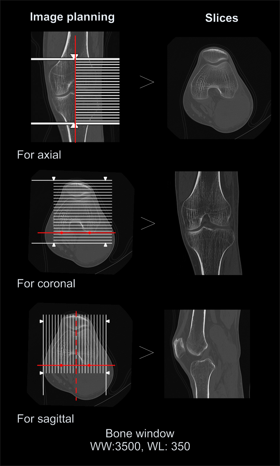
Post-processing
- Axial, sagittal and coronal images in both bone (WW: 3500, WL: 350) and soft tissue window (WW:500, WL:50) with ≤ 2mm slice thickness.
- 3D images to demonstrate pathologies clearly.
Reference
- Mengyuan Kong, Fei Wang, Comparative analysis of computer-aided imaging collaboration: MRI versus CT for detection of knee joint injuries in athletes, Journal of Radiation Research and Applied Sciences, Volume 17, Issue 3, 2024, 100960, ISSN 1687-8507, https://doi.org/10.1016/j.jrras.2024.100960.
- Buzzatti L, Keelson B, van der Voort JW, Segato L, Scheerlinck T, Héréus S, Van Gompel G, Vandemeulebroucke J, De Mey J, Buls N, Cattrysse E, Serrien B. Dynamic CT scanning of the knee: Combining weight bearing with real-time motion acquisition.Knee. 2023 Oct;44:130-141. doi: 10.1016/j.knee.2023.07.014. Epub 2023 Aug 17. PMID: 37597475.
- Chatra P. (2023). The CT knee arthrogram revisited. BJR open, 6(1), tzad007. https://doi.org/10.1093/bjro/tzad007
- Gruenewald LD, Booz C, Martin SS, Mahmoudi S, Yel I, Eichler K, Alizadeh LS, Bernatz S, Gotta J, Reschke P, Weber C, Sommer CM, D'Angelo T, Bucolo G, Leistner DM, Vogl TJ, Koch V. Diagnostic performance of modern computed tomography in cruciate ligament injury detection: A comprehensive study.Eur J Radiol.2024 Jan;170:111235. doi: 10.1016/j.ejrad.2023.111235. Epub 2023 Nov 30. PMID: 38071908.
- Amani Mohamed Fathi El Kharboutly, Multi-detector computed tomography assessment of the tibial plateau fractures, The Egyptian Journal of Radiology and Nuclear Medicine,Volume 46, Issue 3, 2015, Pages 695-699, ISSN 0378-603X, https://doi.org/10.1016/j.ejrnm.2015.05.011.