CT Lumbar spine
Last updated June 27, 2026
By Radiohelp Staff
Similar expressions
CT LS spine/ CT lumbar sacral spine
Introduction
CT LS spine is mainly used to assess lumbar spine injures such as fractures. it also helps to diagnose spine neoplasms, congenital abnormalities and implant checks.
Patient preparation
- Explain the examination clearly and kindly to the patient.
- Patient is asked to remove radio opaque items related to lumbar region.
Patient positioning
- Position the patient in feet-first, supine and in the iso-center.
Explanation: patient in the iso-center reduces radiation exposure and improves image quality.
- Both hands are raised above the head.
Explanation: this reduces streak artifacts due to hands.
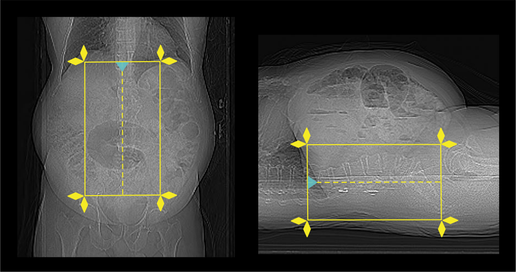
- Plan the scout start point at nipple level and set scan direction outward the gantry.
Scan planning
- Plan the scan slab to cover from T12 to S1.
- Reduce the field of view (FOV) as small as appropriate.
Explanation: smaller FOV increases the geometric resolution of the image.
Post-processing
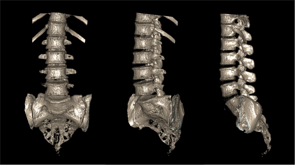
- Sagittal and coronal images with ≤ 2 mm slice thickness in bone window (WW: 3500, WL: 350).
Explanation: curved multi-planar reconstruction (curved MPR) is useful when there are abnormal curvatures in the spine.
- Contagious axial slices in bone window and soft-tissue window (WW:500, WL:50) with ≤ 2mm and ≤ 3mm slice thicknesses respectively.

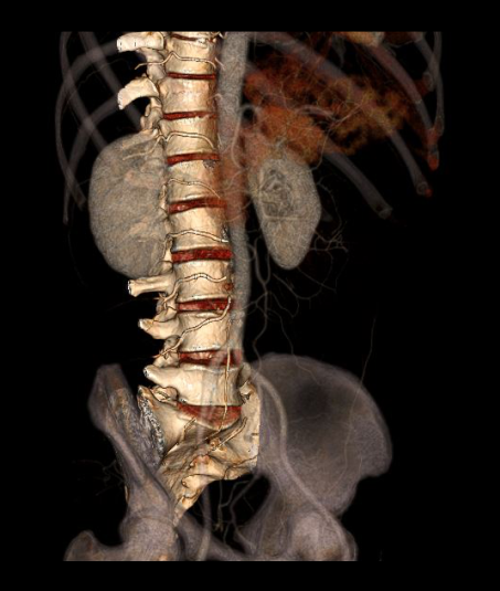
- Additionally, 3d images to show the lumbar-spine.
Reference
- Lubdha M. Shah, MD, Chair, Kristine A. Blackham, MD, & Kavita K. Erickson, MD. (2022). ACR–ASNR–ASSR–SPR practice parameter for the performance of computed tomography (CT) of the spine. Retrieved from www.gravitas.acr.org.
- Ahmad, Z., Mobasheri, R., Das, T., Vaidya, S., Mallik, S., El-Hussainy, M., & Casey, A. (2014). How to interpret computed tomography of the lumbar spine. Annals of the Royal College of Surgeons of England, 96(7), 502–507. https://doi.org/10.1308/rcsann.2014.96.7.502
- Parizel, P. M., van der Zijden, T., Gaudino, S., Spaepen, M., Voormolen, M. H., Venstermans, C., De Belder, F., van den Hauwe, L., & Van Goethem, J. (2010). Trauma of the spine and spinal cord: imaging strategies. European spine journal :official publication of the European Spine Society, the European Spinal Deformity Society, and the European Section of the Cervical Spine Research Society, 19 Suppl 1(Suppl 1), S8–S17. https://doi.org/10.1007/s00586-009-1123-5
- Tins B. Technical aspects of CT imaging of the spine. Insights Imaging.2010 Nov;1(5-6):349-359. doi: 10.1007/s13244-010-0047-2. Epub 2010 Oct 21. PMID: 22347928; PMCID: PMC3259341.
- Lee, S. H., Yun, S. J., Kim, D. H., Jo, H. H., Song, J. G., & Park, Y. S. (2017). Diagnostic usefulness of low-dose lumbar multi-detector CT with iterative reconstruction in trauma patients: a comparison with standard-dose CT.The British journal of radiology, 90(1077), 20170181. https://doi.org/10.1259/bjr.20170181
- Romanyukha, A., Nzitunga, P. S., & Dolcet, A. (2022, April 28). CT patient positioning plays key role in radiation dose reduction.www.auntminnie.com.