CT Venography of lower limbs (direct method)
Last updated August 05, 2026
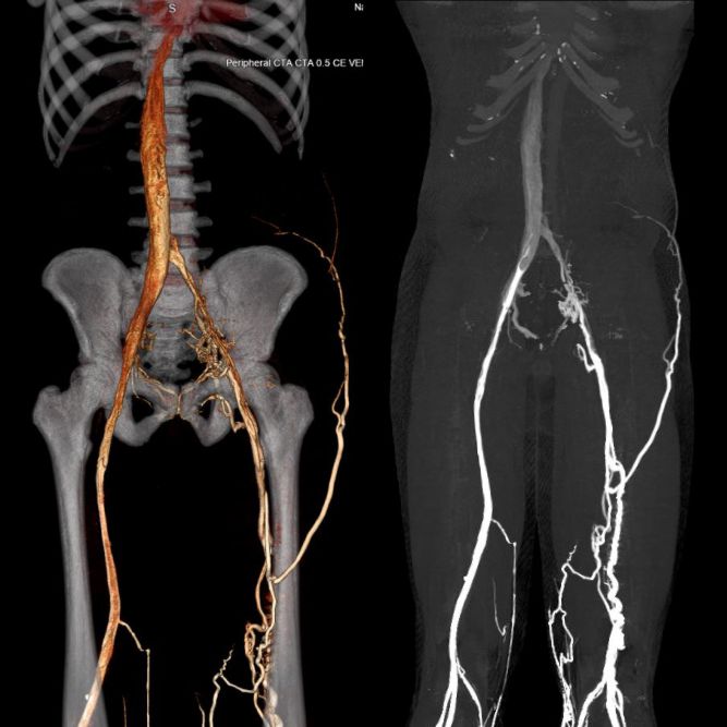
Similar expressions
Please refer to the lower limb CTV indirect method for similar expressions.
Introduction
CT Venogram Lower Limb Protocol is mainly performed using two methods known as the direct and the indirect. These methods differ in the technique of contrast media administration. In the CTV direct method, contrast media is directly injected into the veins of the lower limb, providing good venous enhancement. Whereas in the indirect method, scan is acquired during venous phase after contrast media has drained from organs to veins. Please refer to CTV indirect method for the indications.
Patient preparation
- Assess suitability for radiation exposure and intravenous (IV) iodinated contrast media administration.
- Remove metallic objects in the interested region.
- Remove garments that compress veins such as underwear, stockings, bandages and socks.
- Practice the “breath in and hold” breathing technique.
- Cannulate the lower limb or limbs.
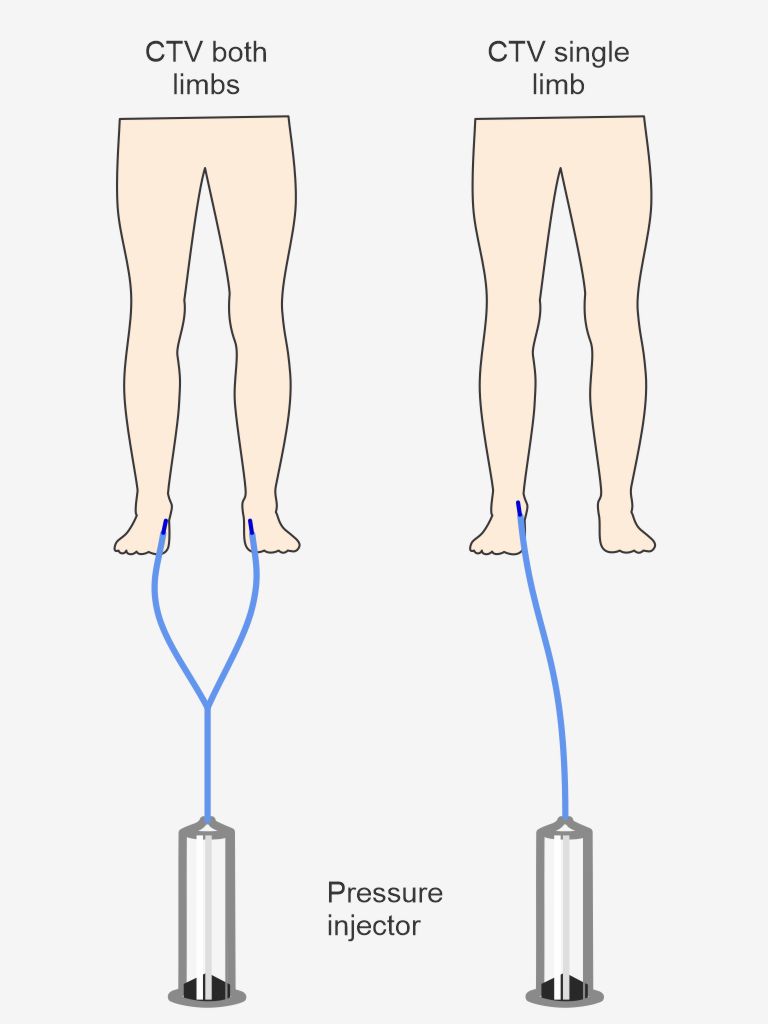
Explanation: proper cannulation of the lower limb is important in this method. Both limbs or a single limb can be cannulated depending on the patient’s clinical indication. A stable superficial vein of a lower limb is cannulated by a professional at the level of the ankle or the foot because veins are enhanced upward from the cannulation point. A 22 gauge-blue cannula is used to inject contrast at a lower contrast flow rate (1.5 ml/s).
Patient positioning
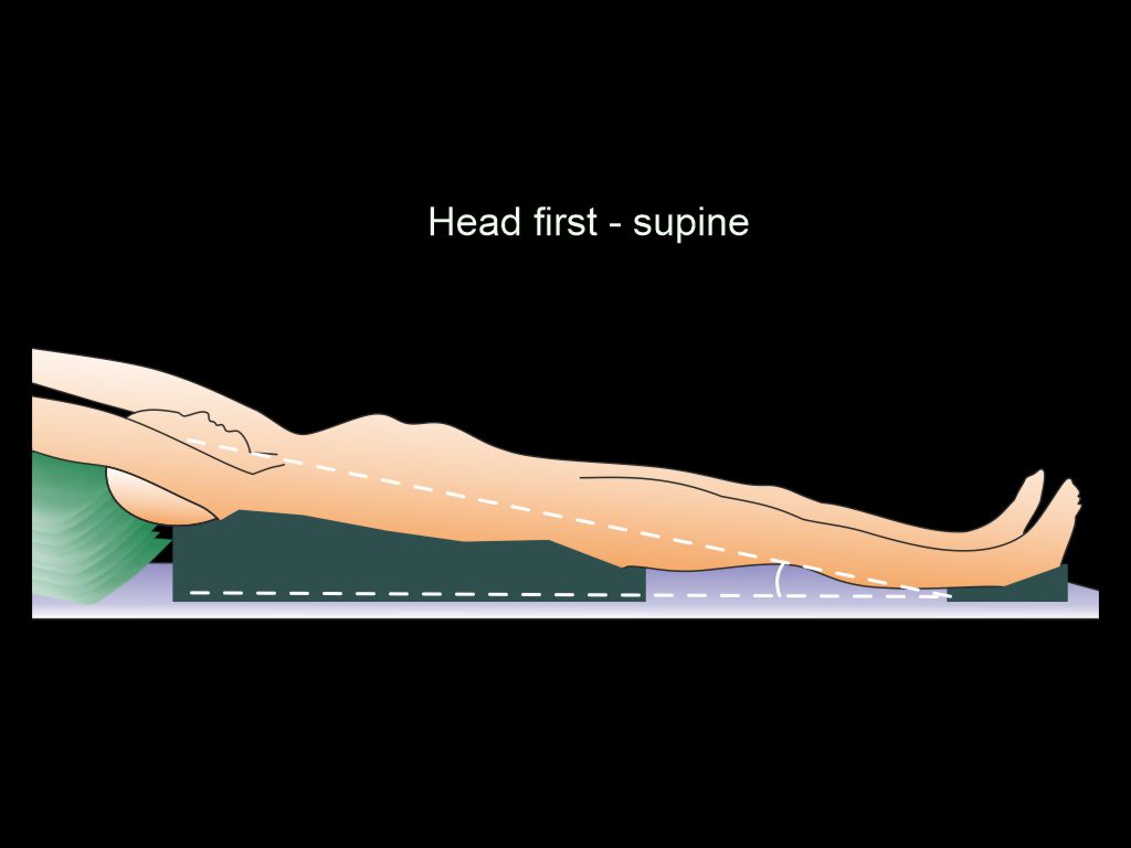
- Position in feet-first supine.
- Place both legs flat on the table.
- Keep padding under the buttocks and heels.
Explanation: reduces compression of the posterior superficial veins caused by the patient’s weight.
- Slightly elevate the pelvis and upper body relative to the heels.
Explanation: delays venous drainage, allowing more time for imaging.
- Keep the region of interest at the scanner iso-center.
Explanation: this reduces the radiation dose to the patient and improves image quality.
- Raise the arms and keep them beside the head.
Explanation: avoids streak artifacts in the chest and the abdominal regions. It also reduces radiation exposure to the patient.
Important: this scan is one of the lengthiest body-coverage CT scans. Therefore, move the couch to check the moving range, and avoid tubing tangling or an accidental stop due to the limited couch range.
Scan planning
The venous phase is acquired using the bolus tracking method.
- Plan the scan slab to cover from the diaphragm to the ends of the lower limbs (toes).
- Set the scan direction from the foot to the head.
Explanation: this helps avoid early image acquisition.
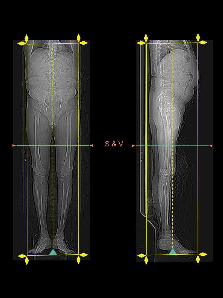
- Keep the Scan and View (S & V) slice at the lower half of the thighs.
- Set no delay between contrast injection to S & V initiation.
- Manually start the venogram when adequate superficial venous enhancement is observed on the S & V scan.
Explanation: automatic triggering cannot be used because the venous anatomy is not clearly visible on the non-contrast S & V slice.
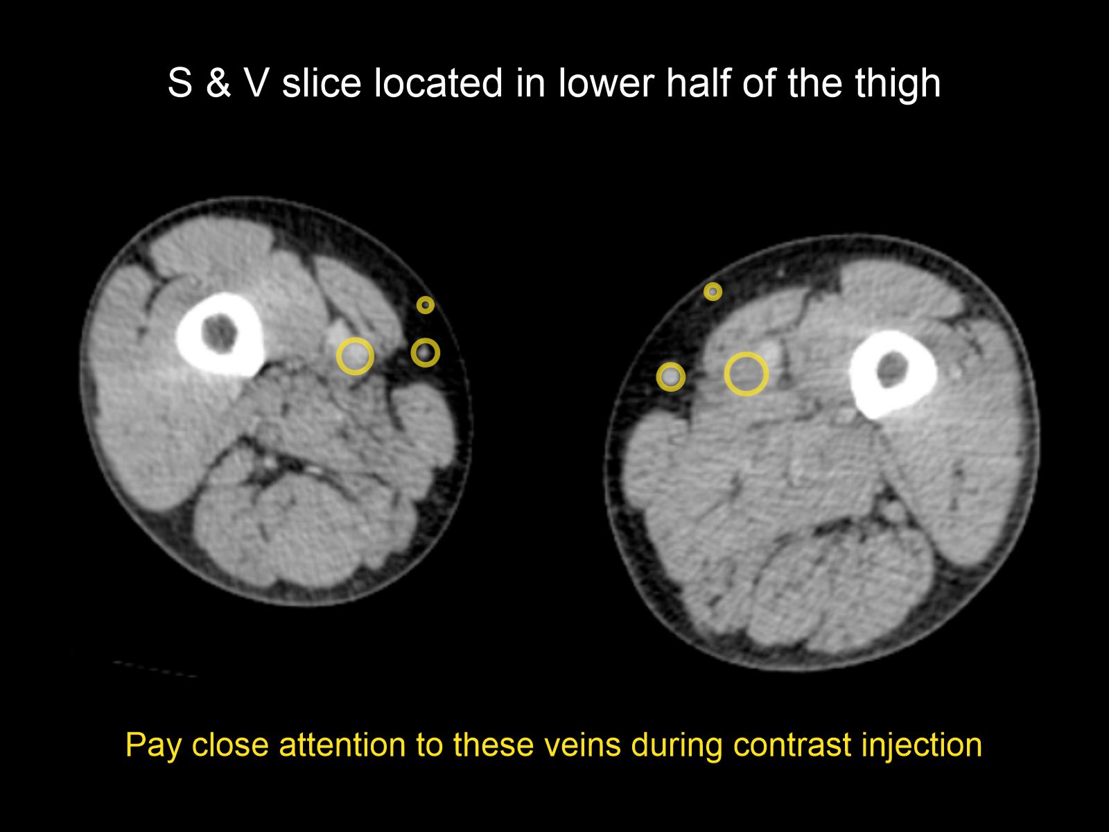
Important: superficial veins are located in the subcutaneous fat. Therefore, attention must be paid to peripheral enhancements in the axial S & V slice.
Intra venous (IV) contrast administration.
Technique and IV contrast volume depend on whether a single limb or both limbs are being imaged. However, IV iodinated contrast need to be diluted and mix well in a 1:3 ratio with one part contrast and three parts normal saline because pure contrast injection results in streak artifacts due to the high density of the contrast medium. For bilateral lower limb CTV imaging, 60-65 ml of contrast mixture is injected simultaneously at a rate of 1.5 ml/s into both limbs via two cannulas, using a Y connector to connect the pressure injector to the cannulas. For single limb CTV imaging, 30-35 ml of contrast mixture is injected at a rate of 1.5 ml/s, and the pressure injector is directly connected to the limb.
Important: IV cannulation of the lower limbs can be challenging due to lower limb swelling. Therefore, the CTV lower-extremity indirect method may be an option if IV cannulation is not possible.
Post-processing
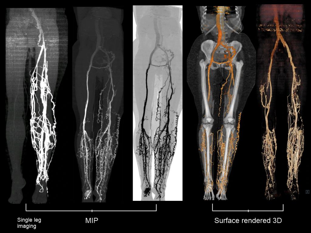
Diagnosis is mainly done using MIP and MPR images. The MIP and surface shaded 3D images of the direct method have better contrast than those obtained with the CTV indirect method because the injected contrast is not absorbed by the organs. Moreover, pre- and post-contrast acquisition for subtraction can also be performed to ease post-processing, but increases patient’s radiation exposure.
- Axial, sagittal and coronal images in both bone (WW: 3500, WL: 350) and soft tissue windows (WW: 500, WL: 50) with ≤ 2 mm slice thickness.
- 3D surface-shaded images and MIP images to demonstrate the lower limb veins.
Reference
- Kim H, Chung JW, Park JH, Yin YH, Park SH, Yoon CJ, Choi YH. Role of CT venography in the diagnosis and treatment of benign thoracic central venous obstruction. Korean J Radiol. 2003 Jul-Sep;4(3):146-52. doi: 10.3348/kjr.2003.4.3.146. PMID: 14530642; PMCID: PMC2698080.
- Mavili E, Ozturk M, Akcali Y, Donmez H, Yikilmaz A, Tokmak TT, Ozcan N. Direct CT venography for evaluation of the lower extremity venous anomalies of Klippel-Trenaunay Syndrome. AJR Am J Roentgenol. 2009 Jun;192(6):W311-6. doi: 10.2214/AJR.08.1151. PMID: 19457795.
- Ana Marija Alduk, M. P. (2018, July). CT Venography: Technique and Indications. Retrieved from www.evtoday.com.
- Sanjit Tewari, MD, Chair, Kevin W. Dickey, MD, FACR, Thomas J. Ward, MD, Christopher D. Yeisley, MD, Mark Baerlocher, MD, & Kush R. Desai, MD. (2023). ACR–SIR Practice parameter for the performance of diagnostic infusion venography. Retrieved from www.gravitas.acr.org