MRI Brain
Last updated August 25, 2026
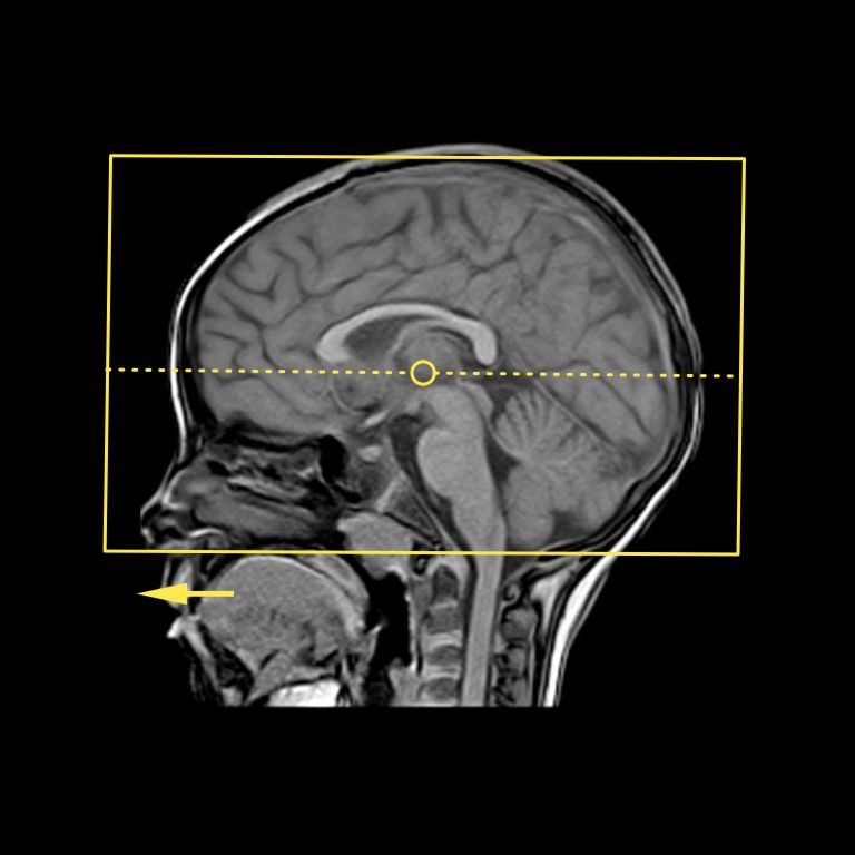
Protocol sequences
| Localizer_3_plane |
| I. T1_Sagittal_Brain |
| II. T2_Axial_Brain |
| III. T2 _Coronal _Brain |
| IV. FLAIR _FS _Axial _Brain |
| V. DWI _Axial _Brain |
| VI. SWI _Axial _Brain |
Introduction
MRI brain scan is one of the routine examinations performed in MR imaging and is used to investigate the causes of symptoms including, but not limited to, headaches, dizziness, tumours, stroke, infections, psychological conditions, and trauma. Post-contrast imaging may be requested to assist in the diagnosis of infections, carcinomas, or for post-surgical assessment. In such cases, gadolinium-based contrast media are administered intravenously.
Patient preparation
- Ask the patient to remove metallic items such as jewellery and remove any clothing that contains ferromagnetic materials and change into a patient gown.
- Obtain MR safety consent after evaluating possible MR contraindications.
- Explain that gradient noise and couch movements are normal.
- Demonstrate how to use the nurse call system and explain when to use it.
- Explain the importance of remaining still during the scan.
- Help claustrophobic patients overcome their fear through demonstrations, or allow a companion to stay with the patient during the scan.
- Inform the patient of the expected duration of the scan.
Patient positioning
- Use the head coil for imaging and position the patient’s head centrally and symmetrically within the coil.
- Place soft pads on either side of the head to immobilize the head and reduce gradient noise.
- Position the patient’s limbs without forming body loops.
Explanation: Body loops may generate eddy currents, which can cause RF burns on the skin. Insulating soft pads may be used to break conductive loops that may form between the feet and thighs or between the trunk and the arm.
- Centre on the glabella.
Sequences in detail
I. T1_Sagittal_Brain
Planning
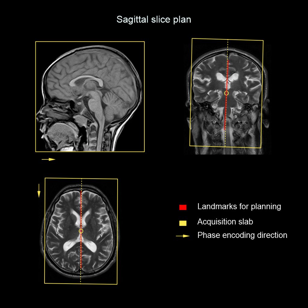
- Scan coverage: From Right temporal lobe to Left.
- Plan block Angulation: Align the slices parallel to the midsagittal plane (MSP) of the brain in both the coronal and axial planes.
Parameters
FOV (cm)24x23
TR530
Matrix192x256
TE11
No Wrap1x1
NAQ2
Slices29
Phase DirectionA to P
Slice thickness (mm)4
FLIP90
Gap (mm)0.8
Purpose
- Anatomical evaluation.
II. T2_Axial_Brain
Planning
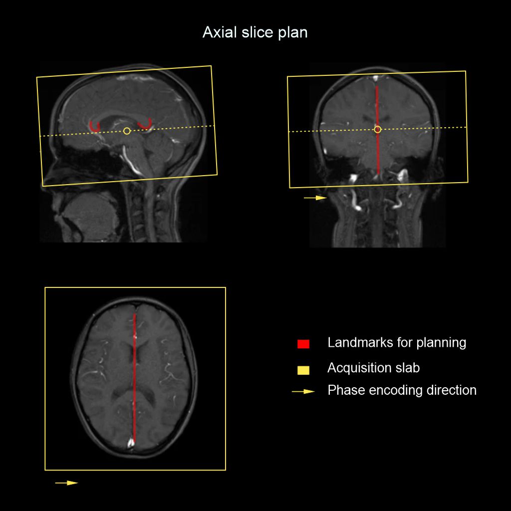
- Scan coverage: Vertex to foramen magnum.
- Plan block Angulation: Align the slices parallel to the line joining the genu and the splenium of the corpus callosum and, in the coronal plane, angle the slices perpendicular to the MSP of the brain.
Parameters
FOV (cm)23x23
TR3800
Matrix256x320
TE90
No Wrap1x1
NAQ1
Slices29
Phase DirectionR to L
Slice thickness (mm)4
FLIP90
Gap (mm)1
Purpose
- Anatomical evaluation.
III. T2_Coronal_Brain
Planning
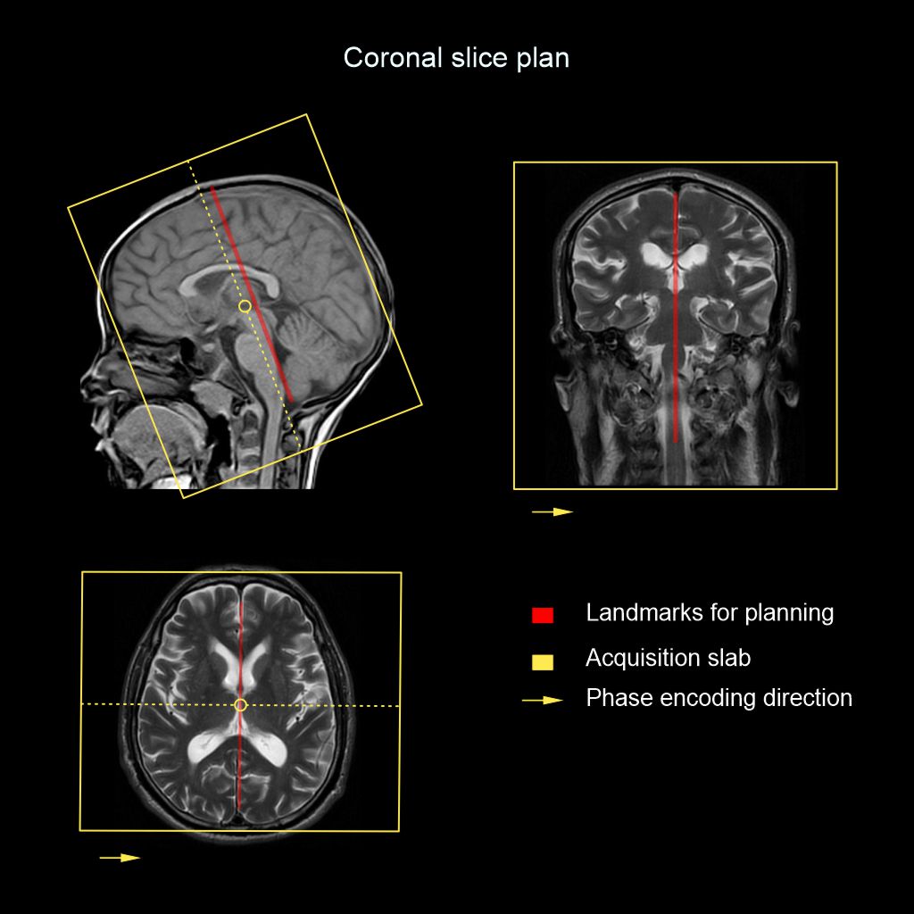
- Scan coverage: From frontal sinus to occipital protuberance.
- Plan block Angulation: Align the slices parallel to the brainstem in the sagittal plane and perpendicular to the MSP in the axial plane.
Parameters
FOV (cm)23x23
TR3800
Matrix320x320
TE90
No Wrap1x1
NAQ1
Slices29
Phase DirectionR to L
Slice thickness (mm)4
FLIP90
Gap (mm)1
Purpose
- Anatomical evaluation.
IV. FLAIR_FS_Axial_Brain
Planning
- Scan coverage: Vertex to foramen magnum.
- Plan block Angulation: Align the slices parallel to the line joining the genu and the splenium of the corpus callosum and, in the coronal plane, angle the slices perpendicular to the MSP of the brain.
Parameters
FOV (cm)23x23
TR11000
Matrix224x224
TE104
No Wrap1x1
NAQ1
Slices29
Phase DirectionL to R
Slice thickness (mm)4
FLIP90
Gap (mm)1
TI2850
Purpose
- Anatomical evaluation (white matter disorders).
V. DWI_Axial_Brain (with ADC)
Planning
- Scan coverage: Vertex to foramen magnum.
- Plan block Angulation: Align the slices parallel to the line joining the genu and the splenium of the corpus callosum and, in the coronal plane, angle the slices perpendicular to the MSP of the brain.
Parameters
FOV (cm)23x23
TR4800
Matrix144x144
TE70
No Wrap1x1
NAQ2
Slices28
Phase DirectionL to R
Slice thickness (mm)4
FLIP90
Gap (mm)1
b Value0, 500, 1000
Purpose
- Assessment of infarction, abscess, dermoid or epidermoid, active demyelination and hypercellular neoplasm.
VI. SWI_Axial_Brain
Planning
- Scan coverage: Vertex to foramen magnum
- Plan block Angulation: Align the slices parallel to the line joining the genu and the splenium of the corpus callosum and, in the coronal plane, angle the slices perpendicular to the MSP of the brain.
Parameters
FOV (cm)23x23
TR29
Matrix256x336
TE20
No Wrap1x1
NAQ1
Slices70
Phase DirectionL to R
Slice thickness (mm)2
FLIP10
Gap (mm)1
Purpose
- Detection of calcifications, microhemorrhages and intravascular thrombosis
Reference
- Eisenmenger LB, Peret A, Roberts GS, Spahic A, Tang C, Kuner AD, Grayev AM, Field AS, Rowley HA, Kennedy TA. Focused Abbreviated Survey MRI Protocols for Brain and Spine Imaging. Radiographics. 2023 Jun;43(6):e220147. doi: 10.1148/rg.220147. PMID: 37167089; PMCID: PMC10262597.
- Saunders DE, Thompson C, Gunny R, Jones R, Cox T, Chong WK. Magnetic resonance imaging protocols for paediatric neuroradiology. Pediatr Radiol. 2007 Aug;37(8):789-97. doi: 10.1007/s00247-007-0462-9. Epub 2007 May 9. PMID: 17487479; PMCID: PMC1950216.
- Mehan WA Jr, González RG, Buchbinder BR, Chen JW, Copen WA, Gupta R, Hirsch JA, Hunter GJ, Hunter S, Johnson JM, Kelly HR, Larvie M, Lev MH, Pomerantz SR, Rapalino O, Rincon S, Romero JM, Schaefer PW, Shah V. Optimal brain MRI protocol for new neurological complaint. PLoS One. 2014 Oct 24;9(10):e110803. doi: 10.1371/journal.pone.0110803. PMID: 25343371; PMCID: PMC4208779.
- Wang C, Shi Z, Li Y, Xia X, Kuang X, Qian S, Xue L, Lan L, Wu Y, Zhang N, Tao J, Hu X, Cao W, He N, Guo Y, Chen W, Zhang J, Luo J, Wang H, Tian M. Protocol for Brain Magnetic Resonance Imaging and Extraction of Imaging-Derived Phenotypes from the China Phenobank Project. Phenomics. 2023 Jan 5;3(6):642-656. doi: 10.1007/s43657-022-00083-w. PMID: 38223689; PMCID:PMC10781909.
- Hutchins, Troy A MD, Chair, Maloney, John A MD, & Srinivasan, Ashok MD. (2024). ACR–ASNR–SPR practice parameter for the performance and interpretation of magnetic resonance imaging (MRI) of the brain. Retrieved from www.gravitas.acr.org.